Debunking myths on genetics and DNA

Showing posts with label neuroscience. Show all posts

Wednesday, March 23, 2016

We Agree to Disagree: The Science of Why Your Political Posts Won’t Make Anyone Change Their Mind


In today's heated political stage, where everyone has a soapbox thanks to outlets like Facebook, Twitter, Instagram and all the personal blogs, I've tried my best not to share my political views publicly. And I've miserably failed. I use my own Facebook page and profile to talk about science, books and photography, but then I can't resist browsing other people's posts. Most of my friends are not as shy as me about making their political views heard and that's when I fall into the trap: I comment. And then someone replies. And I comment back. And on and on it goes until one of us drops out of the conversation because clearly we're not getting anywhere.

Science has taught me to be humble and rational. And yet I'm human, and every time I make a mistake in my line of work I feel something inside my brain stir and protest: "How's that possible? Surely they sent me the wrong data, or they didn't give me the correct information, or the world collapsed and my computer exploded, but there's no way I could've made that stupid mistake."

Apparently, I'm not unique. We all go through this kind of mental distress whenever we encounter an inconsistency between reality and our expectations, and between other people's opinions or choices and our own. It's called "cognitive dissonance." According to Wikipedia, social psychologist Leon Festinger described four ways our brain deals with this:
In an example case where a person has adopted the attitude that they will no longer eat high fat food, but eats a high-fat doughnut, the four methods of reduction are:

  • 1. Change behavior or cognition ("I will not eat any more of this doughnut")
  • 2. Justify behavior or cognition by changing the conflicting cognition ("I'm allowed to cheat every once in a while")
  • 3. Justify behavior or cognition by adding new cognitions ("I'll spend 30 extra minutes at the gym to work this off")
  • 4. Ignore or deny any information that conflicts with existing beliefs ("This doughnut is not high in fat")
What determines what choice we make?

In my case, I end up going back to my computer program. I typically find the bug (which I unknowingly introduced as I was coding), correct it, and rerun the analyses. Admitting my mistake costs me emotional distress, in addition to that nagging doubt at the back of my head -- will my boss still like me even though I made a stupid mistake? -- but in the long run it would cost me a lot more not to correct the error and hand the wrong analyses to our collaborators.

So why can't we do the same when we are heatedly debating politics or religion? Why do some of us even resort to insults rather than admitting that our own logic is faulty?

One possible reason is that there are no consequences to being disrespectful or even offensive when debating on line. After all, even when we use our real name, we are still hiding behind a shield of impersonality when typing our thoughts on an electronic device. On the other hand, if I hand out the wrong results and my collaborators publish them, there will be huge consequences for me. And frankly, trial and error is part of the scientific process: we all make mistakes, we correct them, and we repeat the process over and over again until we have clean and sensible results. Only then we publish a paper.

But in a political or religious debate the consequences can be far more costly if we suddenly admit that we may have been wrong all along. Changing our mind affects our self-esteem and may lead to self-blame, possibly disrupting the relationships around us. That's why our brain has a tendency to choose the easier path, which often coincides with reinvigorating present beliefs rather than shifting to new ones. As Nyhan and Reifler notice in a 2010 paper [1], there's a difference between being uninformed and being misinformed, as the latter is much harder to correct. In the paper, the authors claim that "humans are goal-directed information processors who tend to evaluate information with a directional bias toward reinforcing their pre-existing views," and conclude: "Indeed, in several cases, we find that corrections actually strengthened misperceptions among the most strongly committed subjects."

This behavior of reinforcing one's beliefs the more the contrasting evidence is presented, is called the "confirmation bias". Patterson et al. [2] define this bias as the tendency to favor certain explanations that conform to our own beliefs and/or emotional response, and classify it as "cognitive" or "emotional" depending on whether it reflects the former or the latter. It's a very familiar bias, as we've all seen it everywhere around us, whether it was to defend our favorite presidential candidate or to debate climate change. A little harder is to pin it down when we are engaging in this behavior ourselves -- but rest assured, we all do it at some point, although each one of us to different extents.

"Because of this mechanism," explains Robin S. Cohen, a Los Angeles based psychoanalyst, "not only are we biased to favor perceptions that are in line with our beliefs, but we are also very likely to organize our world in order to only experience things that conform to our own ideas. This makes it less likely to be confronted with alternative opinions. Our own beliefs are so thoroughly reinforced through this process that new perceptions gain very little traction."

Interestingly, as Leonid Perlovsky describes in a 2013 review [3], experiments have shown that music helps abate the stressful consequences of cognitive dissonance. So, maybe I could try playing a little music in the background next time I'm trying to convince a Trump supporter to find a better presidential candidate. What do you think? Mozart or Metallica?

[1] Nyhan, B., & Reifler, J. (2010). When Corrections Fail: The Persistence of Political Misperceptions Political Behavior, 32 (2), 303-330 DOI: 10.1007/s11109-010-9112-2

[2] Patterson, R., Operskalski, J., & Barbey, A. (2015). Motivated explanation Frontiers in Human Neuroscience, 9 DOI: 10.3389/fnhum.2015.00559

[3] Perlovsky, L. (2013). A challenge to human evolution—cognitive dissonance Frontiers in Psychology, 4 DOI: 10.3389/fpsyg.2013.00179

ResearchBlogging.org


Sunday, January 18, 2015

BPA, BPA-free and why Internet titles can be misleading


A few years ago, when I still had both kids in preschool, I became painfully aware that plastic is made of oil. I know, I know, where had I been until then? Underground, I guess. All the bottles I'd used to feed milk and drinks to my kids were scratched and chewed and horrid. I screeched in panic, threw them all away and replaced everything with stainless steel. My kids hated the new bottles and refused to take them to school. Yeah, the joys of parenthood.

So, imagine my joy when BPA-free came around. Finally something my kids will love and that will not harm them. Except, my friend Cristina sent me the bad news a few days ago:
"Dozens of studies link the common plastic chemical bisphenol A (BPA) to all sorts of health problems, including breast and prostate cancers, heart trouble, type 2 diabetes, autism, liver tumors, asthma, infertility, and even obesity. With such a bad track record and hormone-disrupting tendencies, many companies, particularly plastic water bottle manufacturers, have switched to the BPA-free chemical called bisphenol S, or BPS. But a groundbreaking study from University of Calgary researchers suggests we need to diligently avoid both, thanks to newly discovered impacts on the brain [Source]."
Aware of the journalistic exaggerations that infest the Web, I logged onto the PNAS website and downloaded the original paper [1], titled: "Low-dose exposure to bisphenol A and replacement bisphenol S induces precocious hypothalamic neurogenesis in embryonic zebrafish."

Kinch et al. [1] treated embryonic zebrafish (not humans!) with low doses of both BPA and BPS (the compound commonly used in BPA-free products). Apparently, both chemicals are so widespread that, quoting from the PNAS article,
"A recent examination of urine samples in the United States and Asia confirmed previous work showing that 93% of people had detectable levels of BPA but surprisingly showed that 81% had detectable levels of BPS, illustrating the wide-spread use of this poorly known bisphenol analog in consumer products [1]."
BPA molecules behave as receptor antagonists to sex steroid (estrogen or testosterone): what this means is that they bind to the sex steroid receptors, thus blocking the hormone effect. While the general dosage in plastic bottles may be really low to have any effect on adults, the question as to whether or not they are relevant during embryonic development is indeed well posed since that's the time when the hypothalamus is particularly vulnerable due to lack of blood-brain barrier.

The researchers treated the zebrafish embryos with a very low dose for BPA, 1,000-fold lower than the accepted human daily exposure, and then repeated the experiment with the same dosage for BS. They chose the same BPA dose measured in the Oldman River, which serves two major cities in Alberta, Canada. Their results show that BPA exposure in the zebrafish embryos induced hyperactivity and caused precocious neurogenesis in the hypothalamus. Unfortunately, BPS (the BPA-free alternative) wasn't any better, as it still altered brain development and behavior.

Two questions popped in my head. First, the researchers immersed the embryos in the contaminants, whereas human embryos are immersed in amniotic fluid, which is always filtered by the placenta, a filter between what circulates in the mother's blood stream and the child's. Given this, did Kinch et al. make a fair comparison? The researchers address the placenta issue, claiming that BPA concentration has been measured in the human placenta and that the dosage used in the experiment was 100-fold lower than circulating levels found in fetal serum.

The other question a critical reading of the paper should pose is: how good of a model is the zebrafish embryo for human embryonic development? Especially knowing that zebrafish embryos develop in a mere 72 hours and grow up to a couple of centimeter long, whereas the hypothalamus in the human embryo starts forming around week 8, when it's roughly 1.5 centimeters. It turns out, it is a good model when it comes to embryonic neurogenesis:
"Despite the large evolutionary distance between fish and mammals, the overall organization, basic structures, and functional capacities of major hypothalamic components are highly conserved between zebrafish and mammalian brains. [...] In contrast to mammals, zebrafish embryos develop externally and are transparent, and highly amenable to genetic manipulation, making them an ideal vertebrate model for in vivo studies of neural patterning and neuronal specification. As such, zebrafish models have been used extensively in recent years to study the roles played by key signaling pathways in controlling the development of hypothalamic neurons [2]."
Having addressed in a satisfactory way my two main concerns with the PNAS paper [1], I particularly like the two points the researchers make in the discussion section. The first one is that the way tolerable levels are determined is using a linear model, starting from the highest possible dose and gradually lowering until no effect is detected. Kinch et al. rightfully argue that dose-response relationships are hardly ever linear when it comes to endocrine-disrupting compounds like BPA and BPS.

Second, the researchers claim that the switch from BPA to BPS was done without adequate toxicology testing. What if it turns out that BPS is comparable to BPA in terms of damaging potential?

BPA and BPS aside, I wanted this post to also make a case for critical thinking. I'm seeing way too much junk on the Internet being passed for science when in fact it's just bad reporting/journalism (in genetics in particular!!). While the Internet article my friend sent me didn't make any incorrect statement, and it did discuss the actual study a few paragraphs into the article, still, the title, "Popular 'BPA-Free' Chemical Causes Brain Damage, Study Finds" had me frowning because it lacks to mention two fundamental points: 1) the study was conducted on zebrafish (not on humans!) and 2) the supposed brain damage was on the zebrafish embryos (not on humans, and not on children or adults).

In fact, let me open a quick parenthesis and cite some numbers a friend of mine posted on Facebook: a paper on some break-through research on Alzheimer's published by the Journal of Clinical Investigation titled "Prostaglandin signaling suppresses beneficial microglial function in Alzheimer’s disease..." got shared 116 times. Stanford posted the story under the title "Blocking receptor in brain’s immune cells counters Alzheimer’s in mice, study finds" and got shared almost 10,000 times. The Telegraph titled its piece "Has Stanford University found a cure for Alzheimer's disease?" and got almost 50,000 shares. But at the very top of the sharing contest came Rod D. Martin  (over 130,000 shares) with the title "Happy New Year! Stanford May Have Just Cured Alzheimer’s" .

Bottom line 1: yeah, you can get very popular when you blow up science.
Bottom line 2: if you want to draw some useful conclusions from any pseudo-sceintifc news, go back to the source before getting too excited and/or too alarmed.

The zebrafish experiment indicates that compounds containing BPA and BPS could potentially harm the fetus and as a safety precaution it recommends pregnant women to steer away from plastic bottles, but it doesn't present any evidence on the effects on children or (non-pregnant) adults. It does not conclude that BPA and/or BPS exposure has the same effect in humans, but it does pose a foundation for further studies as it raises a flag that there may be a risk associated to these chemicals. The way to further validate this hypothesis would be to measure BPA and BPS levels in the urine of pregnant women and then follow the babies' neurological development for a few years to see if the women with higher levels had problematic children compared to the women with lower levels.

Of course, it's still a good idea to avoid plastic and use it only when other alternatives aren't handy. Plastic use has certainly increased exponentially in the past few decades and it's a good idea to reduce its use anyways, since it could not only be potentially harmful for our bodies, but it's also definitely bad for the environment. As in all things, cautiousness (whether it's used to avoid plastic or to avoid Internet content) is the best measure for a happy living.

 [1] Kinch, C., Ibhazehiebo, K., Jeong, J., Habibi, H., & Kurrasch, D. (2015). Low-dose exposure to bisphenol A and replacement bisphenol S induces precocious hypothalamic neurogenesis in embryonic zebrafish Proceedings of the National Academy of Sciences DOI: 10.1073/pnas.1417731112

 [2] Machluf, Y., Gutnick, A., & Levkowitz, G. (2011). Development of the zebrafish hypothalamus Annals of the New York Academy of Sciences, 1220 (1), 93-105 DOI: 10.1111/j.1749-6632.2010.05945.x

ResearchBlogging.org

Sunday, January 11, 2015

The viruses inside us: can endogenous retroviruses elicit antibodies?

January Moonrise © EEG

Today I would like to discuss a couple of papers that I used as premise for my new thriller Immunity, which will be part of the Apocalypse Weird series, created by Nick Cole, Michael Bunker and Tim Grahl. Just like all my other thrillers, Immunity too, finds its roots in some fascinating facts about genetics, virology and of course immunity.

The premise of the book has to do with something I discussed a long time ago, in one of my very first posts: human endogenous retroviruses, or HERV's, are small portions of our DNA that we acquired from ancient retroviruses that infected germ line cells of our primate ancestors. Basically, these genes came directly from retroviruses that inserted themselves into cells that then became oocytes or spermatozoa and, once fertilized, passed the viral genes to a new individual. These genomic elements are mostly inactivated in adults (meaning they are in a non-coding part of the DNA), but they have been shown to be transcriptionally active during fetal development. The intriguing bit, however, is that expression levels of these genetic elements have been found to be disrupted in subjects with schizophrenia [1].

I'm sure you are all familiar with the disease, which typically manifests itself through hallucinations (mostly auditory ones), delusions, and the inability to distinguish reality from things that only exists in the patient's mind. It's often characterized also by disorganized thoughts and incoherent speech. Nobel laureate John Nash suffered from schizophrenia, and his disease was portrayed in the movie A Beautiful Mind, though in a very fictionalized way. Another famous case is USC professor Elyn Saks, who wrote an award-winning memoir on her life-long battle against schizophrenia.

Retroviruses are sleek little things. They can infect brain cells and integrate their genomes into the host cell's DNA, causing all sorts of damage. For example, some studies have shown that viruses like HIV and HTLV can indeed infect the brain, causing symptoms such as psychosis and depression [2]. The body fights viruses and pathogens by sending its sentinels (natural killer cells, T cells and antibodies) to find them and destroy them. But what happens if the virus is already embedded in our genome, as is the case with HERVs? Those viral elements have been part of our genome for millions of years, so, in theory, our immune system is not supposed to 'see' them.

 One of the most marvelous and yet most delicate mechanisms that is at the foundation of our immune system is its ability to distinguish self from non-self. T cells and B cells have to undergo strict scrutiny to make sure that they don't mistakenly attack cells of our own body thinking that they are pathogens. This mechanism is tough but not perfect, and failures to recognize self from non-self are at the basis of numerous auto-immune disorders. Autoimmune thyroditis, for example, is an inflammation of the thyroid caused by antibodies attacking the thyroid.

One natural hypothesis as to why HERVs expression levels could be disrupted in a disease like schizophrenia could be that the body is producing antibodies against those genetic elements. This hypothesis cannot be tested directly because, as Dickerson et al. explain in [1], there are no available reagents. However, one can look for antibodies that recognize retroviruses like murine leukemia virus (MuLV), Mason-Pfizer monkey virus (MPMV), and feline immunodeficiency virus (FIV) because they have enough similarities with HERVs.

Dickerson et al. measured the levels of antibodies against these viruses in a population of 666 study subjects, of which 163 with a recent onset of psychosis, 268 with multi-episode schizophrenia, not of recent onset, and 235 controls without a history of psychiatric disorders. They found a significant increase in antibody levels in the recent onset group compared to controls, but not in the multi-episode group compared to controls. At the same time, these subjects had no traces of the actual viruses in their bodies, indicating that the antibody response had to be elicited by the endogenous elements (instead of an active infection). Another study [2] looked for an enzyme called reverse transcriptase, which is a marker for retroviral activity, and found that it was 4 times higher in the cerebrospinal fluid of patients with recent onset of schizophrenia compared to controls.

Many autoimmune disorders are caused by the immune system suddenly attacking its own self. I've used this premise before in my books: Track Presius, the main character in Chimeras, has elevated levels of anti-nuclear antibodies, which are antibodies that, in high concentrations, can cause different immunological disorders as they tend to bind to human antigens.

What intrigued me about the HERV-schizophrenia association, though, was: the researchers tested the presence of antibodies against HERV's using viruses that are not commonly found. What if, instead, a common virus like the flu did bear resemblance to the HERV elements in our brain? In order to fight the infection, our body would have to start producing antibodies that could potentially attack those human genes, too. What would then happen to the brain, suddenly under attack by its own antibodies?

I don't know the real answer, but I can tell you that I had fun speculating about it in my novel. Immunity will be released in April and it will be part of the Apocalypse Weird series.

[1] Dickerson F, Lillehoj E, Stallings C, Wiley M, Origoni A, Vaughan C, Khushalani S, Sabunciyan S, & Yolken R (2012). Antibodies to retroviruses in recent onset psychosis and multi-episode schizophrenia. Schizophrenia research, 138 (2-3), 198-205 PMID: 22542615

[2] Yolken R (2004). Viruses and schizophrenia: a focus on herpes simplex virus. Herpes : the journal of the IHMF, 11 Suppl 2 PMID: 15319094

ResearchBlogging.org

Sunday, November 2, 2014

The heritability of fears

Cyborg © EEG
As many of you know, one of my favorite topics here on the blog is epigenetic inheritance, i.e. the mechanisms that regulate changes in gene expression that can be passed from one generation to the next. Epigenetics has revolutionized the way we look at genetic inheritance: Darwin had taught us that the only way the environment can shape the genome of a species is through natural selection. While this is certainly still true, today we also know that:

1) Most of the mutations we see in a population have reached fixation through random drift -- the constant reshuffling from one generation to the next -- not selection.

2) The environment can induce changes in one generation that may indeed be passed on to the next generation not through actual changes in the DNA but, rather, in the way the DNA is "packaged" inside the cell nucleus (for a great explanation on how this work, see my colleague Karissa Sanbomatsu's TED talk).

 In a Nature Neuroscience paper [1], authors Dias and Ressler explored the following premise in a mouse model:
"An important, but often ignored, factor that influences adult nervous systems is exposure of parents to salient environmental stimuli before the conception of their offspring. Such information transfer would be an efficient way for parents to ‘inform’ their offspring about the importance of specific environmental features that they are likely to encounter in their future environments. However, this would necessitate the transgenerational inheritance of environmental information via the germ line by offspring not even conceived at the time."
The researchers used olfactory fear as the stimulus mostly because it's one of the best understood mechanisms, both at the neurological and the molecular biology levels. Of course, a caveat would be that humans, besides being very different from mouse models, they've evolutionarily replaced olfactory stimuli with visual ones.

The researchers used odor-naive male mice and targeted an odorant receptor (M71) whose expression in the olfactory sensory neurons has been shown to be activated by acetophenone. It is important to note that the experiment did not induce any change in the actual DNA of the mice. What they did, instead, was use acetophenone to activate the receptor so it would be expressed inside these special neurons.

As I explained in older posts, DNA is wrapped around "spools" called histones. Cells produce proteins and activate receptors depending on what genes are on the outer surface of the "histone yarn", while hidden parts of the DNA remain unexpressed (as if that gene didn't exist). A molecule like acetophenone can induce changes inside olfactory sensory neurons that cause the histones to move and expose the gene that encodes the M71 odorant receptor. Once this happens, the receptor is "activated."

 Since the mice are initially odor naive, their M71 receptor is inactivated (the gene is not expressed) prior to the exposure to acetophenone. After the receptor activation, these male mice were mated with odor naive females. So, genetically speaking, the offsprings had no reason to have the M71 receptor activated, since neither parent had it activated at birth. Yet the offsprings of the mice stimulated with acetophenone, despite not being previously exposed to any of the odors with which they were tested, were able to detect acetophenone at lower concentrations than the offsprings of mice stimulated with another molecule (propanole).

Not all offsprings were behaviorally tested. Some of the offsprings were kept naive to any exposure so that their neuroanatomy could be tested separately without risking the results to be affected by the behavioral tests. When they looked at the offsprings of the acetophenone exposed mice, the researchers found an increase in the M71 glomerular area together with a significant increase in the numbers of M71-activated olfactory sensory neurons in the main olfactory epithelium.

So, how these epigenetic changes get inherited? To address the question, Dias and Ressler examined the sperm of the acetophenone exposed mice. This part of the paper gets a little technical, but the interesting idea is that they did find molecular changes in the sperm DNA around the Olfr151 gene, which encodes the M71 receptor. They found that the 3' end of Olfr151 was significantly less methylated in the acetophenone induce mice. At the same time, they
"did not observe any histone-mediated epigenetic signatures around the M71 locus when chromatin was immunoprecipitated with antibodies that recognize histone modifications that either permit or repress to transcription."
The authors conclude:
"In summary, we have begun to explore an under-appreciated influence on adult behavior—ancestral experience before conception. From a translational perspective, our results allow us to appreciate how the experiences of a parent, before even conceiving offspring, markedly influence both structure and function in the nervous system of subsequent generations. Such a phenomenon may contribute to the etiology and potential intergenerational transmission of risk for neuropsychiatric disorders, such as phobias, anxiety and post-traumatic stress disorder."

[1] Dias, B., & Ressler, K. (2013). Parental olfactory experience influences behavior and neural structure in subsequent generations Nature Neuroscience, 17 (1), 89-96 DOI: 10.1038/nn.3594

ResearchBlogging.org


Friday, May 9, 2014

Prying minds with mind-blowing optogenetics


Did you know there was such a thing as optogenetics? The idea alone completely blows my mind:
"Optogenetics uses light to control neurons which have been genetically sensitised to light. It is a neuromodulation technique employed in neuroscience that uses a combination of techniques from optics and genetics to control and monitor the activities of individual neurons in living tissue to precisely measure the effects of those manipulations in real-time. The key reagents used in optogenetics are light-sensitive proteins." [Wikipedia]
The "light sensitive proteins" mentioned above are a family of proteins, called opsins, that are found in the photoreceptor cells of the retina. These proteins are responsible for converting light (photons) into electrochemical signals.

So, in layman terms, the idea behind optogenetics is that if we can deliver these opsin proteins into the neurons, making them sensitive to light, we can then use light to control the neurons themselves. This is used to understand the function of certain cell types in the brain. How do you deliver the proteins to the neurons? Using viral vectors, of course. When injected into the brain, the viral vectors infect the neurons, delivering the opsin genes. These genes make the neurons sensitive to light and can therefore be activated or silenced using optical fibers delivering light. It sounds very much like science fiction, but basically this enables researchers to control neurons using optical fibers.

Source: Lumencor
This optic stimulation is limited to very small areas of the brain. Not only that. The way neurons react to light depends on the frequency used to stimulate them. Animal studies have shown that light stimulation of the ventral segmental area can induce depressive-like behaviors at 20 Hz, whereas increasing to 30 Hz (in a different study) elicited antidepressant effects.

Because there's a whole family of opsin proteins, current research is aimed at understanding which ones work best depending on the experimental setting and circumstances. For example, different opsins can elicit neurons at different wave lengths, and when there's no overlap between the two spectra, two different opsin proteins can be used simultaneously to obtain two different outcomes on neural activity. Pushing this even further, genes coding for these proteins can be mutated to change their wave-length and frequency sensitivity and can be optimized for certain experimental settings.

Researchers use optogenetics to identify brain circuits that control emotions like fear, depression, and anxiety, and all the areas involved in those circuits. Previous methods included local lesions, pharmacological treatment, and electrophysiological studies, but these didn't give complete control on the temporal window like light stimulation does, which can activate or inhibit neurons at a very precise moment. It's fascinating stuff that I confess I don't completely understand myself as it is not my field, so I welcome the input from any experts out there willing to share their view and any literature recommendations!

On a side note, CHIMERAS is now at $.99 for a limited time only! (Grab a copy if you love mysteries and science).

[1] Belzung C, Turiault M, & Griebel G (2014). Optogenetics to study the circuits of fear- and depression-like behaviors: A critical analysis. Pharmacology, biochemistry, and behavior, 122C, 144-157 PMID: 24727401

ResearchBlogging.org

Sunday, March 30, 2014

What do one trillion different scents smell like?


I've been really happy with the comments on my upcoming detective thriller CHIMERAS. The book will be released in two weeks, but I've already heard back from some early readers (and yes, I'm still offering free ARC's, see details here), and many have praised Track's sensitivity to smells. Apparently, it's a trait many relate to and yet you don't find so often in fiction.

Most of our memories are stored as images. So, even when we write, we tend to over-emphasize visual descriptions and forget all about our nose. From a scientific point of view, though, how does olfaction compare to other senses? Can we "see" and "hear" more than we can "smell"?
"Humans can discriminate several million different colors and almost half a million different tones, but the number of discriminable olfactory stimuli remains unknown. The lay and scientific literature typically claims that humans can discriminate 10,000 odors, but this number has never been empirically validated [1]."
In a study published last week in Science, Bushdid et al. calculated that, contrary to previous estimates, humans can discriminate at least one trillion different olfactory stimuli -- far more than colors and tones.

How did they make such an estimate?

Colors are created from light: changes in wavelength create different hues and saturations. Similarly, sounds are created from air waves and changes in frequencies create different tones. Because we can physically measure both the frequency and wavelength of waves, it is relatively easy to determine the ranges within which human eyes and ears can detect these stimuli:
"Humans can detect light with a wavelength between 390 and 700 nm and tones in the frequency range between 20 and 20,000 Hz [1]."
But while colors and tones are created by waves, olfactory stimuli are created by mixtures of numerous distinct odor molecules. Even the "simple" scent of a rose contains 275 components. The odor molecules bind to the olfactory receptor cells in the nasal cavity, sending a signal up to the olfactory nerve.

One trivia that I discovered while writing CHIMERAS is that olfactory receptors are not restricted to the nasal cavity. They are also found in sperm cells [2] where they are possibly involved in the control of sperm migration and fertilization.

To measure the resolution of the human visual or auditory system, scientists measure how close two signals need to be in frequency in order to become undistinguishable. In other words, if the signals are like hair and our ability to pick them up is a comb, how fine are the comb teeth? How far apart do two light wavelengths need to be in order for our eyes to discern them as distinct colors?

Bushdid et al. used a similar criterion to measure how good we are discerning scents. They used 128 odor molecules to make different scent mixtures of 10, 20, or 30 components. Each mixture yields a different smell, and the more components the mixtures shares the harder they are to distinguish from one another. So, similarly to what's typically done for the visual and auditory system, in order to measure the resolution of the human nose, Bushdid et al. measured how much two mixtures need to overlap in order to become indistinguishable to the human nose. Of the 26 subjects in the study:
"At least half of the tested subjects could discriminate mixture pairs that overlapped by less than 75% of their components. Some could also discriminate mixture pairs that overlapped by 75 and 90%, but none could discriminate mixture pairs with more than 90% overlap [1]."
Bushdid et al. then used mathematical extrapolations to predict that the majority of individuals can distinguish mixtures that overlap less than 51%, which amounts to over one trillion mixtures made with 30 components. This is only a lower limit since for their experiment the researchers used 128 different components while in nature you can find many more, and in mixtures of often more than just 30 components.

I wonder what they would've concluded had Track been part of the study. :-)

[1] Bushdid C, Magnasco MO, Vosshall LB, & Keller A (2014). Humans can discriminate more than 1 trillion olfactory stimuli. Science (New York, N.Y.), 343 (6177), 1370-2 PMID: 24653035

[2] Vanderhaeghen P, Schurmans S, Vassart G, & Parmentier M (1993). Olfactory receptors are displayed on dog mature sperm cells. The Journal of cell biology, 123 (6 Pt 1), 1441-52 PMID: 8253843

ResearchBlogging.org

Saturday, December 14, 2013

ASD and inflammation: more than just a correlation


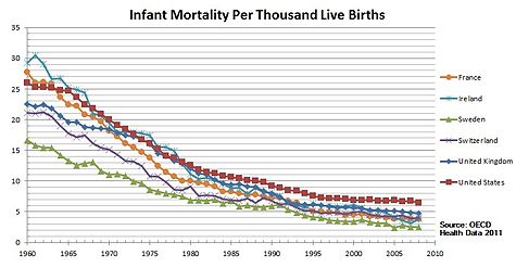
There has been a lot of speculation, lately, about vaccines possibly being harmful and, in particular, causing autism. You know I work on HIV vaccine design, so there's no need to say where I stand on the need of vaccinations. No link has been found between the incidence of autism and vaccination. Of course, medicine is not an exact science. Outliers will always exist. The U.S. seem to be a special case, as the vaccination schedule in this country requires a high number of vaccine doses, yet the infant mortality rate is one of the highest among North America and European countries. However, take a close look at this graph:


The countries with low mortality rate shown in this graph have a strict vaccination schedule, just like the U.S. On the other hand, what distinguishes them from the US is affordable health care. Countries with a high infant mortality rate are countries where poor people do not have access to vaccines and good health care. For the 3-million AIDS orphans living in sub-Saharan Africa a vaccine against HIV is the only hope they have to live into adulthood. It is quite easy for those of us who have a healthy life style and have access to food, medicines, and doctors on a daily basis, to say "no, thank you" to vaccines. But please, when you make your own decision about vaccines, do remember the millions of people for whom this is not a choice. And also remember: some children who are immunodefecient really cannot be vaccinated. They cannot contract any kind of disease, either, because their immune system is not working. However, if the majority of the people continue to get vaccinated, people who really cannot be vaccinated are still protected:

found on Facebook

Back to autism. As you saw from my last post, ASD, or autism spectrum disorders, is indeed a puzzling disease and pinning down its etiology has been challenging. The genetics involve numerous genes and diverse pathways, implying that different mechanisms could potentially lead to ASD, particularly during fetal development. One thing that I recently discovered is a number of correlations found between infections in the mother during gestation and autism:
"Recent studies have highlighted a connection between infection during pregnancy and the increased risk of autism in the offspring. Parallel studies of cerebral spinal fluid, blood and postmortem brains reveal an ongoing, hyper-responsive inflammatory-like state in many young as well as adult autism subjects. There are also indications of gastrointestinal problems in at least a subset of autistic children [1]."
In his review [1], Patterson makes a good summary of the relevant studies: for example, a permanent, inflammatory-like state has been found in postmortem examination of ASD affected brains. This was found at all ages, indicating that the state was established early in the development and maintained throughout the life-span of the ASD affected individual. These abnormalities expand to the central nervous system and the peripheral immune system affecting also the gastrointestinal tract:
"These findings include immune cell infiltrates present in the colon, ileum and duodenum, as well as increased T cell activation in the intestinal mucosa. These inflammatory changes are associated with autoimmune responses that could contribute to the observations of decreased mucosal integrity, or 'leaky gut' [1]."
"Abnormal activation of the immune system may also be involved in the etiology of autism. [. . .] Family members of autistic children, particularly the mothers, show a higher incidence of allergy or autoimmune diseases. Consistent with immune involvement are findings that maternal infection is a risk factor for autism [2]."
In conclusion, there is a correlation between immune abnormalities and ASD, and the immune abnormalities propagate to the brain and the gastrointestinal tract. However, it is unclear if these abnormalities cause the behavioral symptoms of ASD or if they are a secondary effect. The health and well-being of our immune system has such deep, profound effects on the central nervous system. The two interact very closely together: stress and the general emotional status, for example, can affect immunity; vice versa, the immune system can influence behavior. Both our brain and our immune system constantly learn and readapt to the surrounding environment (for example, our immune system learns to recognize new pathogens throughout our lifetime), which makes them prone to life-long epigenetic changes induced by environmental factors such as stress and disease. It's not a coincidence that:
"Immune dysregulation has also been implicated in the etiology of a variety of neurodegenerative, psychiatric, and neurodevelopmental disorders, including Parkinson, Huntington, and Alzheimer diseases, multiple sclerosis, major depression, schizophrenia, and addiction [2]."
Hsiao et al. [2] addressed the open question of whether the immunological abnormalities cause ASD-like behaviors in a mouse model. They induced ASD in mouse offspring through "maternal immune activation" (MIA): the immune system of pregnant mice was altered and then the offsprings of the altered mice that were behaviorally abnormal was compared to the offsprings of the controls. The behaviorally abnormal MIA offsprings exhibited core behavioral symptoms of autism, including increased repetitive behaviors, decreased social interactions, and increased anxiety. Hsiao et al. found several abnormalities in the immune system of these MIA offsprings: levels of regulatory T-cells were decreased and CD4+ T-cells were hyper-responsive. These abnormalities could not be transferred to healthy mice through a bone marrow from the MIA mice. However, when irradiated and transplanted with immunologically normal bone marrow, many of the behavioral abnormalities stopped. This would suggest that the immunological dysregulation causes the ASD-like behaviors.
"It is striking that in a mouse model of an autism environmental risk factor that exhibits the cardinal behavioral and neuropathological symptoms of autism, there is also permanent peripheral immune dysregulation. This finding provides the opportunity to explore molecular mechanisms underlying the relationship between brain dysfunction and altered immunity in the manifestation of abnormal behavior. Furthermore, this finding provides a platform for investigating how prenatal challenges can program long-term postnatal immunity, health, and disease. Maternal insult-mediated epigenetic modification in HSC and progenitor cells is one possible mechanism for how effects may be established by transient environmental changes yet persist permanently into adulthood. However, the BM transplant results suggest that the peripheral environment of the MIA offspring is also critical for maintaining a permanently modified immune state [2]."
We will never be able to prove or disprove a direct causal relation between vaccines and autism: if a child develops ASD after vaccination, unfortunately, we cannot rewind time and see if the same child, without the vaccine, would've never developed ASD in his/her lifetime. ASD typically develops in infancy, which is when the bulk of vaccines are administered. The risk of ASD is much higher (see last week's post) if there's already a family member with ASD, siblings in particular. And given the deep, complex interactions and reciprocal influence between the nervous system and the immune system it is quite possible that a sudden change in the immune system could cause some level of disruption in the nervous system. However, if the immune system is primed to such risk, a virus or any other pathogen, which cause changes in the immune system just like a vaccine does, could also cause similar disruptions. On the other hand, vaccines can potentially prevent infections that, according to these studies, do increase the risk of ASD in the baby during the first trimester of gestation.

So, as always: Read the literature, talk to your doctor, possibly to more than one, consider your family's medical history, and, whatever decision you make, make sure it is an informed decision.

[1] Patterson, PH (2011). Maternal infection and immune involvement in autism Trends in Molecular Medicine DOI: 10.1016/j.molmed.2011.03.001

[2] Hsiao EY, McBride SW, Chow J, Mazmanian SK, & Patterson PH (2012). Modeling an autism risk factor in mice leads to permanent immune dysregulation. Proceedings of the National Academy of Sciences of the United States of America, 109 (31), 12776-81 PMID: 22802640

ResearchBlogging.org

Sunday, December 8, 2013

Autism: not one disease but a spectrum of disorders; not one gene but a network of gene coexpressions.


"Autism spectrum disorder (ASD) is a lifelong developmental condition that affects about 1 in 110 individuals, with onset before the age of three years. It is characterized by abnormalities in communication, impaired social function, repetitive behaviors and restricted interests [1]."
ASD is more common among males than females, with a 4:1 male to female ratio. Numerous studies in the literature have shown evidence for a strong genetic component of autism, with a risk up to 25 times higher among siblings compared to the general population. However, if you look at the literature, you find that these numbers change pretty dramatically from study to study. This is often the case when you look at rare disorders in conjunction with rare mutations (WARNING: the rest of the paragraph is a statistical digression, feel free to skip to the next section). The smaller the effect you are trying to measure, the more subjects you will need in your study. This is also true if you are testing many variants, as for example in GWAS studies, which investigate variants in the whole genome. If the effect is big enough, you will find statistical support for your association, however, if your sample size is not big enough, the effect you are trying to measure will vary greatly from study to study. This is because the smaller the sample size, the larger the variance, which is stat jargon to say that whatever you are trying to measure (typically an increase in risk) is likely to be different if you repeat the study.

What do we know about the genetic etiology of ASD? About 10% of people diagnosed with ASD have some underlying genetic syndrome (including mitochondrial genes). About 5% are due to rare chromosome rearrangements, for example changes in the size, shape, or number of some chromosomes. Another 5% has been associated to both inherited and de novo "copy number variations" (CNV), the presence of extra copies of some genes [1]. CNV is not rare among humans, as it accounts for approximately 0.4% of the variation between unrelated genomes. Identical twins also differ in CNV, and, even though they have identical genomes, the copy number of the genes may differ between the two. Despite this, in some families with a history of ASD the proportion of de novo CNV's has been found to be up to five times higher than in families without a history of ASD. Finally, thanks to recent advances in sequencing technology, de novo point mutations throughout hundreds of genes have been found and implicated in about 15% of ASD cases [2].

In light of the variety of mutations, genes, and phenotypes associated with ASD, two studies published in the last issue of Cell addressed the following question:
"do these genetic loci converge on specific biological processes, and where does the phenotypic specificity of ASD arise, given its genetic overlap with intellectual disability (ID)? [2]"
"if and when, in what brain regions, and in which cell types specific groups of ASD-related mutations converge during human brain development [3]" ?
Of the two papers, I've so far only read the one by Willsey et al. [3], who combined their own data with already published data and identified 144 de novo "loss-of-function (LoF)" mutations, in other words, mutations that impair the functionality of the gene (hence the corresponding protein is no longer produced). They called genes with 2 or more de novo LoF mutations "hcASD", or "high confidence" ASD because statistically they had a high probability of being truly associated with ASD. They also analyzed a less-likely set of genes with only one de novo LoF mutation, which they called "pASD genes".

Next, the researchers investigated when and where these genes are expressed during brain development. The way they did this is a bit technical, but to think about it in simple terms think of it this way: (1) they needed samples from brain tissues taken at different developmental stages; (2) they needed to look not just at one gene, but at families of genes that are likely to interact together and influence one another's likelihood of getting turned "on" and "off". When a gene is turned "on", the gene is coding a protein, and we say that the gene is "expressed."

To carry on their analysis, Willsey et al. used data published by Kang et al. (Nature, 2011) from "57 clinically unremarkable postmortem brains of diverse ancestry (31 males, 26 females) that span 15 consecutive periods of neurodevelopment and adulthood from 5.7 postconceptual weeks (PCW) to 82 years." The gene expression values were determined for each gene by brain region and by postmortem brain sample. Brain regions were grouped according to transcriptional similarity during fetal development. These data were used to generate 52 gene coexpression networks, each network composed of the hcASD genes and their top correlated genes. This coexpression network analysis is a technique that's been extensively used lately to analyze patterns of co-expressions of genes. Each gene in the network is represented by a node, and any two nodes (genes) at any given time are connected if the genes are expressed at that time.

Using this set-up, the researchers were able to link the ASD genes to particular brain regions and developmental phases.
"Our analysis identifies robust, statistically significant evidence for convergence of the input set of hcASD and pASD risk genes in glutamatergic projection neurons in layers 5 and 6 of human midfetal prefrontal and primary motor-somatosensory cortex (PFC-MSC). Given the extensive genetic and phenotypic heterogeneity underlying ASD and the small fraction of risk genes that we have examined in this study, this likely represents only one of several such points of convergence. Nonetheless, the analytic approach presented here clarifies key variables relevant for productive functional studies of specific ASD genes carrying LoF mutations, providing an important step in moving from gene discovery to an actionable understanding of ASD biology [3]."
Cortical glutamatergic projection neurons (CPNs) are a class of neocortical neurons. They are called "projection" neurons because they transmit information from the neocortex to other neocortical and central nervous system regions. During development, projection neurons are generated in the neocortical germinal zone and migrate radially to their final neocortical position. In their study, Wyllsey et al found that the development of midfetal CPNs is particularly vulnerable to ASD. Furthermore, the set of ASD genes they identified as associated to ASD are functionally diverse and encode proteins found in distinct cell compartments, confirming the theory that ASD can be caused by different and distinct pathways.
"Given recent studies suggesting that as many as 1,000 genes or more could contribute to ASD (He et al., 2013; Iossifov et al., 2012; Sanders et al., 2012), our analysis has uncovered a surprising degree of developmental convergence. Despite starting with only nine hcASD seed genes, we have identified highly significant and robust evidence for the contribution of coexpression networks relevant to L5 and L6 CPNs in two overlapping periods of midfetal human development (3–5 and 4–6) corresponding to 10–24 PCW [3]."
The importance of these studies lies in the understanding of not just the genetic association per se, but in the mechanisms that drive these associations, and, most importantly, how the numerous genes interact and when.

[1] Devlin and Schrer (2012). Genetic architecture in autism spectrum disorder Genetics & Development DOI: 10.1016/j.gde.2012.03.002

[2] Neelroop N. Parikshak, Rui Luo, Alice Zhang, Hyejung Won, Jennifer K. Lowe, Vijayendran Chandran, Steve Horvath, Daniel H. Geschwind (2013). Integrative Functional Genomic Analyses Implicate Specific Molecular Pathways and Circuits in Autism Cell DOI: 10.1016/j.cell.2013.10.031

[3] A. Jeremy Willsey, Stephan J. Sanders, Mingfeng Li, Shan Dong, Andrew T. Tebbenkamp, Rebecca A. Muhle, Steven K. Reilly, Leon Lin, Sofia Fertuzinhos, Jeremy A. Miller, Michael T. Murtha, Candace Bichsel, Wei Niu, Justin Cotney, A. Gulhan Ercan-Sencicek, J (2013). Coexpression Networks Implicate Human Midfetal Deep Cortical Projection Neurons in the Pathogenesis of Autism Cell DOI: 10.1016/j.cell.2013.10.020

ResearchBlogging.org

Friday, December 7, 2012

The simulated brain


His name is Spaun, which stands for Semantic Pointer Architecture Unified Network, and he's a brain -- a simulated, brain. His 2.5 million neurons, organized in subsystems that simulate different brain areas, allow Spaun to perform tasks such as image recognition and recalling sequences, and respond through a motor arm. For example, Spaun can recognize numbers on a screen and write them on a piece of paper.

Spaun is the brain child (pun intended!) of authors Eliasmith et al. [1]. It models three specific brain areas: the prefrontal cortex for memory, the basal ganglia to select actions, and the thalamus. Spaun's functional architecture consists of a working memory that, given a visual input, compresses the information and translates the input into firing patterns. The next step is the action selection step, which results in a motor output through the robotic arm. Spaun's memory doesn't just store information, but it also correlates new information with the old one. A nice feature of the model is that different neuron parameters can be chosen from random distributions in order to simulate different population behaviors. This simulates the human brain so well that Spaun expresses a common human behavior: the tendency to remember best the first and last items in a list.

On the other hand, Spaun exhibits noteworthy deviations from human brains: while it can get better and better at a particular task, it cannot learn a completely new task. Another shortcoming is that Spaun's attention and eye position are fixed, so that, contrary to a real human brain, it cannot control the input.

As the authors explain:
"Anatomically, many areas of the brain are missing from the model. Those that are included have too few neurons and perform only a subset of functions found in their respective areas. Physiologically, the variability of spiking in the model is not always reflective of the variability observed in real brains. However, we believe that, as available computa- tional power increases, many of these limitations can be overcome via the same methods as those used to construct Spaun."

[1] Eliasmith, C., Stewart, T., Choo, X., Bekolay, T., DeWolf, T., Tang, Y., & Rasmussen, D. (2012). A Large-Scale Model of the Functioning Brain Science, 338 (6111), 1202-1205 DOI: 10.1126/science.1225266

ResearchBlogging.org


Thursday, March 15, 2012

Young or old it doesn't matter: we need them both


To honor Brain Awareness Week I thought I'd try and discuss a neuroscience paper this week. It's not my field, so you'll have to be patient with me (and you experts out there are more than welcome to pitch in). I found a really fascinating story in the latest issue of Science [1] on the differences in information processing between "young" and "old" neurons. In order to understand the story, I had to take a couple of steps back and review a few things about the brain.

The hippocampus is the part of the brain that's responsible for learning, storing memories and associating them with feelings and emotions. Within the hippocampus lies the dentate gyrus, which is where adult neurogenesis takes place -- the formation of new neurons throughout adulthood. The middle layer of the dentate gyrus contains a type of neurons called granule cells. These are constantly generated and take a few weeks to develop and integrate in the dentate gyrus network. In [1], Marin-Burgin et al. asked the following question:
"Is it solely the continuous addition of new neurons to the network that is important, or are there specific functional properties only attributable to new granule cells (GCs) that are relevant to information processing?"
In order to answer the question, the researchers compared immature granule cells to mature ones in mouse hippocampus. The part that fascinates me the most about these experiments is that in order to "see" the different cells, these neurons are "retrovirally labeled to express red fluorescent protein." What this means is that a genetically engineered retrovirus that preferentially infects this type of cells is used to "infect" them and deliver the fluorescent proteins so that the neural activity can be visualized. Pretty cool, right?

Marin-Burgin et al. found that the dentate gyrus is made of a heterogeneous population of granule cells of different ages and that the different subpopulations have distinct activation thresholds. When given both excitatory and inhibitory input, the ratio of excitation to inhibition favors inhibition in mature granule cells, whereas immature cells have fewer inhibitory inputs (hehe, sounds familiar don't you think?). In other words, younger cells respond more easily and broadly, whereas older cells tend to be more specific. The fact that both are present at all times suggests that this range in different responses is needed for the correct functionality of the dentate gyrus, in particular for the correct storing and integration of novel information.

[1] Marin-Burgin, A., Mongiat, L., Pardi, M., & Schinder, A. (2012). Unique Processing During a Period of High Excitation/Inhibition Balance in Adult-Born Neurons Science, 335 (6073), 1238-1242 DOI: 10.1126/science.1214956

ResearchBlogging.org

Tuesday, January 3, 2012

A lot happens in the blink of an eye!


Do you have recurrent nightmares? I do, especially when I'm under a lot of stress. I often dream of missing a train. The setting, location, company and place I need to reach change every time, but the common factor is always the frightening sense of having missed the train and not being able to make it. Another recurrent nightmare I have is that the light is so bright I can't keep my eyes open. So I start blinking faster and faster but I can't see a thing and end up missing something very important.

Blinking seems to be such an important task that our body takes care of constantly, as important and essential as breathing. And yet we hardly ever think about it. Under normal conditions, we blink spontaneously 10 to 15 times a minute. We blink both eyes at the same time, which may seem obvious but (maybe you already knew this, I didn't!) it's unique to mammals. Birds, for example, blink one eye at the time and this prevents loss of visual information. In fact, blinking causes a momentary loss of vision that lasts lasts 100 to 150 milliseconds -- a mini black-out that happens constantly as we stare at things and yet we hardly ever notice it, in ourselves, or in others.

Another thing our eyes smoothly cope with is saccades, quick, simultaneous movements of both eyes. When I rapidly move my camera and press the shutter, not matter how fast the shutter speed, I get a blur. However, our eyes move all the time and yet what we perceive is a constant, flowing image.

We compensate these visual disruptions with two similar mechanisms: blinking suppression and saccadic suppression -- basically, visual sensitivity is suppressed immediately prior to and during both blinking and saccades. The two mechanisms are often coordinated in order to minimize downtime in visual processing. Unfortunately,
They are challenging to study because any brain-activity changes resulting from an extra-retinal signal associated with the blink motor command are potentially masked by profound neural-activity changes caused by the retinal-illumination reduction that results from occlusion of the pupil by the eyelid [1].
In order to measure the neural consequences of blinks on visual function one has to bypass the physical consequences of eyelid closure. In other words, you want to stimulate the retinae maintaining the eyes open. How? Via the mouth, as Volkmann et al. [2] showed in 1980: you insert a light probe in the mouth, the light passes through the palate and stimulates the retinae without forcing eyelid closure.

In [1], Bristow et al. use the same technique to stimulate both retinae while measuring brain activity through fMRI. They also used opaque goggles to ensure that retinal illumination remained constant throughout the experiment, whether the eyelids were open or closed. By doing this, they could see what parts of the brain were responding to the retinal stimulation independently from the change in illumination caused by eyelid closure. They find that
Whereas it might have been supposed that blink suppression is a purely low-level visual phenomenon, mediated solely by retinotopic visual areas, our whole-brain analysis surprisingly revealed that activity evoked by retinal stimulation in parietal and frontal cortices was also suppressed by blinking.

Thus, one possible interpretation of our findings is that the observed suppression of these parietal and prefrontal regions during blinking represents a neural mechanism underlying the lack of awareness of the changes in visual input that normally occur during a blink. Specifically, it may account for the lack of awareness of the percept of the eyelid descending across the pupil and the resulting reduction in retinal illumination.
Their experiment also proves the deep connection between saccade suppression and blinking suppression, as they conclude:
In summary, our data demonstrate that responses to retinal illumination are suppressed by blinking in retino-topic visual area V3 and in parietal and prefrontal cortices, whereas in the absence of retinal stimulation, we identified a positive blink-related signal in early visual areas LGN–V3. We propose that these findings represent a neural signature of blinking associated with the blink motor command and may go some way toward explaining both the neural mechanisms underlying the visual-sensitivity loss, known as blink suppression, that occurs during blinks, and why they go unnoticed. Our findings parallel recent observations of saccade-related changes in activity in visual cortex during saccades, suggesting that blink suppression and saccadic suppression may indeed share common neural mechanisms.
Finally, I'd like to mention a more recent paper by Bonfiglio et al. [3], which used EEGs to look at brain waves during blinking. Brain EEGs typically show oscillations that are classified based on their frequency. Spontaneous blinking modulates two oscillations in particular, alpha and delta, which are thought to be involved in the automatic mechanism of maintaining visual awareness. In their study, Bonfiglio et al. studied the alpha oscillations and postulated that
a) an early blink-related synchronization represents the short-term memory maintenance of the last visually perceived trace of the surroundings; b) the alpha blink-related desynchronization is associated with the comparison between the newly perceived image of the environment and its mnestic representation.

[1] Bristow, D., Haynes, J., Sylvester, R., Frith, C., & Rees, G. (2005). Blinking Suppresses the Neural Response to Unchanging Retinal Stimulation Current Biology, 15 (14), 1296-1300 DOI: 10.1016/j.cub.2005.06.025

[2] Volkmann FC, Riggs LA, & Moore RK (1980). Eyeblinks and visual suppression. Science (New York, N.Y.), 207 (4433), 900-2 PMID: 7355270

[3] Bonfiglio L, Sello S, Carboncini MC, Arrighi P, Andre P, & Rossi B (2011). Reciprocal dynamics of EEG alpha and delta oscillations during spontaneous blinking at rest: a survey on a default mode-based visuo-spatial awareness. International journal of psychophysiology : official journal of the International Organization of Psychophysiology, 80 (1), 44-53 PMID: 21238505

Photo: trees in downtown illuminated over the holidays. It was freezing cold, but the sky was so pretty and I just couldn't stop shooting. I could hardly feel my hands when I got home.

ResearchBlogging.org