Debunking myths on genetics and DNA

Showing posts with label genetic effects. Show all posts

Friday, April 29, 2016

Hunting For The Signatures of Cancer

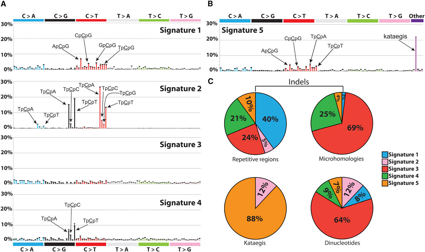
Signatures of Mutational Processes Extracted from the Mutational Catalogs of 21 Breast Cancer Genomes. Credit:http://dx.doi.org/10.1016/j.celrep.2012.12.008

Cancer is the second leading cause of death worldwide, with approximately 14 million new cases and 8.2 million cancer related deaths each year (Source: WHO). A family history of cancer typically increases a person's risk of developing the disease, yet most cancer cases have no family history at all. This suggests that a combination of both genetics and environmental exposures contribute to the etiology of cancer. In this context, "genetics" means the genetic make-up we are born with and inherited from our parents. For example, women born with specific mutations in the BRCA1 and BRCA2 genes are known to have a much higher risk of developing breast cancer later in life.

However, besides the genetic make-up we carry from birth, there are many geographical and environmental factors that contribute to the risk of cancer. For example, the incidence of breast cancer is over 4 times higher in North and West Europe compared to Asia and Africa (Source: WHO). Stomach cancer, on the other hand, is much more prevalent in Asia than the US. If you think that this may be linked to the genetic differences across ethnicities, think again. The National Cancer Institute published a summary of several studies that compared the incidence of first and second generation immigrants in the US with the local population. They found that:
"cancer incidence patterns among first-generation immigrants were nearly identical to those of their native country, but through subsequent generations, these patterns evolved to resemble those found in the United States. This was true especially for cancers related to hormones, such as breast, prostate, and ovarian cancer and neoplasms of the uterine corpus and cancers attributable to westernized diets, such as colorectal malignancies."
According to the World Health Organization (WHO),
"around one third of cancer deaths are due to the 5 leading behavioral and dietary risks: high body mass index, low fruit and vegetable intake, lack of physical activity, tobacco use, alcohol use."
Cancer is the result of a series of cellular mechanisms gone awry: every time a cell divides, somatic mutations accumulate in the cell's genome. These are not mutations we are born with, inherited from our parents. Rather, these are changes that accumulate in certain cells as we grow old and are not  the same across all cells in the body. Many environmental exposures contribute to this process and affect the rate at which these mutations accumulate. However, cells have various mechanisms that are normally able to repair harmful mutations or, when the damage is beyond repair, to trigger cell death. The immune system is also "trained" to recognize cancer cells and destroy them.

When all these defense mechanisms fail, cancer cells start dividing uncontrollably.

As a result, all cancer cells carry a number of somatic mutations that set them apart from healthy cells, and some tend to be the same across different cancer patients: for example, specific mutational patterns found in lung cancer have been attributed to tobacco exposure and were indeed reproduced in animal models. Another set of mutations has been attributed to UV exposure and has been found in skin cancers [1, 2].

This prompts the ambitious question: can we find common mutations across individuals with the same cancer? And how many of these mutational patterns that are common across individuals can we attribute to particular exposures and/or biological processes? Distinguished postdoctoral researcher Ludmil Alexandrov, from the Los Alamos National Laboratory, has been working on this problem since his he was a PhD student at the Wellcome Trust Sanger Institute.

"It's like lifting fingerprints," Alexandrov explains. "The mutations are the fingerprints, but now we have to do the investigative work and find the 'perpetrator', i.e., the carcinogens that caused them." During his graduate studies, under the supervision of Mike Stratton of the Wellcome Trust Sanger Institute, Alexandrov developed a mathematical model that, given the cancer genomes from a number of patients, is able to pick the "common signals" across the patients -- i.e. mutation patterns that are common across the patients -- and classify them into "signatures."

"When formulated mathematically," Alexandrov explains, "the question can be expressed as the classic 'cocktail party' problem, where multiple people in a room are speaking simultaneously while several microphones placed at different locations are recording the conversations. Each microphone captures a combination of all sounds and the problem is to identify the individual conversations from all the recordings." Taking from this analogy, each cancer genome is a "recording", and the task of the mathematical model is to reconstruct each conversation, in other words, the mutational patterns. These are sets of somatic mutations that are the observed across the cancer genomes and that characterize certain types of cancers.

In 2013, Alexandrov and colleagues analyzed 4,938,362 mutations from 7,042 patients, spanning 30 different cancers, and extracted more than 20 distinct mutational signatures [2]. "Some patterns were expected, like the known ones caused by tobacco and UV light," Alexandrov says. "Others were completely new."

Of the new signatures found, many are involved in defective DNA repair mechanisms, suggesting that drugs targeting these specific mechanisms may benefit cancers exhibiting these signatures [3]. But the most exciting part of this research will be finding the 'perpetrator' or, as Alexandrov explains, the mutations triggered by carcinogens like tobacco, UV radiation, obesity, and so on. The challenge will be to experimentally associate these mutational patterns to the exposures that caused them. In order to do this, the scientists will have to expose cultured cells and model organisms to known carcinogens and then analyze the genomes of the experimentally induced cancers.

In the meantime, the signatures found so far are only the beginning: Alexandrov and colleagues have teamed up with the Los Alamos High Performance Computing Organization in order to analyze the genomes of almost 30,000 cancer patients.

"The amount of data we will have to handle for this task is enormous, on the order of petabytes," Alexandrov says. "Few places in the world have the capability to handle this many data. Under normal circumstances, it takes months to answer a question on 10 petabytes of data. The supercomputing facility at Los Alamos can provide an answer within a day."

Because of his research, in 2014 Alexandrov was listed by Forbes magazine as one of the “30 brightest stars under the age of 30” in the field of Science and Healthcare. In 2015 he was awarded the AAAS Science & SciLifeLab Prize for Young Scientists in the category Genomics and Proteomics [2] and the Weintraub Award for Graduate Research. He is now the recipient of the prestigious Oppenheimer fellowship at Los Alamos National Laboratory.

References
Siegel, R., Miller, K., & Jemal, A. (2015). Cancer statistics, 2015 CA: A Cancer Journal for Clinicians, 65 (1), 5-29 DOI: 10.3322/caac.21254

[1] Alexandrov LB (2015). Understanding the origins of human cancer. Science (New York, N.Y.), 350 (6265) PMID: 26785464

[2] Alexandrov LB, Nik-Zainal S, Wedge DC, Aparicio SA, Behjati S, Biankin AV, Bignell GR, Bolli N, Borg A, Børresen-Dale AL, Boyault S, Burkhardt B, Butler AP, Caldas C, Davies HR, Desmedt C, Eils R, Eyfjörd JE, Foekens JA, Greaves M, Hosoda F, Hutter B, Ilicic T, Imbeaud S, Imielinski M, Jäger N, Jones DT, Jones D, Knappskog S, Kool M, Lakhani SR, López-Otín C, Martin S, Munshi NC, Nakamura H, Northcott PA, Pajic M, Papaemmanuil E, Paradiso A, Pearson JV, Puente XS, Raine K, Ramakrishna M, Richardson AL, Richter J, Rosenstiel P, Schlesner M, Schumacher TN, Span PN, Teague JW, Totoki Y, Tutt AN, Valdés-Mas R, van Buuren MM, van 't Veer L, Vincent-Salomon A, Waddell N, Yates LR, Australian Pancreatic Cancer Genome Initiative, ICGC Breast Cancer Consortium, ICGC MMML-Seq Consortium, ICGC PedBrain, Zucman-Rossi J, Futreal PA, McDermott U, Lichter P, Meyerson M, Grimmond SM, Siebert R, Campo E, Shibata T, Pfister SM, Campbell PJ, & Stratton MR (2013). Signatures of mutational processes in human cancer. Nature, 500 (7463), 415-21 PMID: 23945592

[3] Alexandrov LB, Nik-Zainal S, Siu HC, Leung SY, & Stratton MR (2015). A mutational signature in gastric cancer suggests therapeutic strategies. Nature communications, 6 PMID: 26511885

ResearchBlogging.org

Saturday, December 12, 2015

Hunting for the signatures of cancer


Today I'm proud to introduce you to a talented postdoctoral fellow in my own group at the Los Alamos National Laboratory. No, I had nothing to do with his work, which is why I can discuss it without any competing interests. Ours is the Theoretical Biology group, and what that means is that we do biology from a purely theoretical perspective: we design analytical models and analyze data from experiments. Sounds trivial, but it's not, and it takes the joint forces of people coming from the most disparate fields to do what we do: in our group, you'll find physicists, immunologists, biologists, statisticians, mathematicians, and then more physicists.

The particular research I want to discuss today involves looking at the genomes of cancer cells. Perhaps the most famous mutations associated with cancer are the ones found in the genes BRCA1 and BRCA2. These are germline mutations, i.e. mutations that are found in certain people from birth. Women who have these mutations in the BRCA1 and/or BRCA2 genes have a 60% higher chance of developing breast cancer during their lifetime than those who don't (and yet 80% of breast cancers are not associated to these mutations, see this older post for more details on that).

DNA has a certain likelihood to accumulate new random mutations every time the cell divides. These are called somatic mutations, i.e. mutations that aren't present at birth, but arise as we age. Some environmental exposures like smoking and radiation can also cause somatic mutations. Cancer tissue, as you can imagine, is riddled with somatic mutations, but, as it turns out, the mutations differ from cancer to cancer, and also depending on what exposure caused the disease. For example, certain mutational patterns occur most frequently in lung cancer caused by smoking, while others in skin cancers caused by ultraviolet light. These mutational patterns become "signatures" of a particular cancer, and the question is: can we predict the prognosis of the disease based on these signatures? Can we find specific treatments that work for specific signatures?

Some drugs are already in use that work only with cancer tissues that have specific receptors.

Ludmil Alexandrov, a postdoc in my group, used the incredible wealth of DNA sequencing data from tumor tissues to develop a mathematical framework that analyzes the different mutational patterns of each single cell genomes and explore how these signatures developed over time. As Alexandrov wrote in his recent article [1] in Science:
"I curated the majority of publicly available data and compiled a data set encompassing ~5 million somatic mutations from the mutational catalogs of 7042 primary cancers of 30 different classes. These data revealed the existence of 21 distinct mutational signatures in human cancer. Some were present in many cancer types, [. . .] others were confined to a single cancer class."
Because of this work, which he developed during his graduate research at the Wellcome Trust Sanger Institute, Ludmil won the Science and SciLifeLab prize for young scientists (awarded by the American Association for the Advancement of Science and Science Magazine) and the 2015 Weintraub Award for Graduate Research. In his own words,
"In summary, my Ph.D. thesis provided a basis for deciphering mutational signatures from cancer genomics data and developed the first comprehensive census of mutational signatures in human cancer. The results reveal the diversity of mutational processes underlying the development of cancer and have far-reaching implications for understanding cancer etiology, as well as for developing cancer prevention strategies and novel targeted cancer therapies."
Congratulations, Ludmil, well deserved!

[1] Alexandrov, L. (2015). Understanding the origins of human cancer Science, 350 (6265), 1175-1177 DOI: 10.1126/science.aad7363

ResearchBlogging.org

Sunday, February 15, 2015

Brain mosaicism and altered gene copy numbers could explain Alzheimer's

© EEG

I've tackled the problem of the missing heritability in the past, i.e. the fact that despite all the research on genetic studies and disease associations, we can explain only a small fraction of cancers and disorders. Today we know a lot more than what we knew back when the human genome project was completed, and in particular we know how much we don't know. I think we are only beginning to understand the complexity of human diseases and genetics. Back when I started working on disease associations, in 2004, we roughly thought that we could find a few "buttons" that would trigger cancer. Today we know that it's not about finding a few buttons. We thought DNA was more or less a keyboard, when in fact we have a whole orchestra: DNA, mRNA, proteome, epigenome, etc. Mutations can occur at any level, and besides genetic alterations there can be epigenetic alterations, proteins that don't fold correctly, an abnormal accumulation of proteins, and so many other ways that things can go wrong. To this add exercise, body mass, diet, and all kinds of other environmental exposures.

Bottom line: we set out looking for a few "keys" to play on the DNA keyboard when in fact we should be looking for a whole symphony, and the symphony may very well change from person to person.

Alzheimer's disease is among the many disorders that have baffled researchers. Recent studies have found that rather than genetic mutations in the DNA we should be looking at abnormal accumulation of misfolded proteins called amyloids. They can accumulate inside cells to a level where they become toxic and cause cell death. Amyloids have been associated with many diseases, not just Alzhemeir's, but in the case of Alzheimer's in particular they seem to be responsible for the progressive loss of neurons and brain connections. A gene called APP codes for a protein that is an amyloid precursor, and mutations in this gene have been associated with familial Alzheimer's (when the disease occurs before age 60). However, the vast majority of Alzheimer's cases occur much later in life and are not associated with those mutations. Do all these cases fall into yet another missing heritability mystery?

As it turns out, genetic alterations come in many forms, not just mutations. The key, in the case of APP, could be not in what kind of gene allele one carries, but rather on how many copies we carry. Yes, we may have more than two copies in different parts of the brain.

Remember when they taught us in school that we are born with one DNA and that DNA is identical throughout our cells and tissues? Forget that. Of all organs, the brain is one of the most plastic regions of our body, with numerous retrotransposons and mobile genetic elements that attest for its plasticity. Errors in chromosome segregation when cells divide can also produce cells with a gain or loss in chromosome numbers. This variability has been shown to be a common feature of the normal brain: our brains are genetic mosaics made of genetically distinct cell lines that have, over the years, accumulated somatic mutations [1].

I've discussed retrotransposons a while back: these are genetic elements that can make copies of themselves and then reinsert in different parts of the DNA. They are particularly active in the brain and thanks to their activity the brain is in fact a somatic mosaic of genetically distinct neurons. Retrotransposons were first discovered in maize and explain why, for example, a single cob can have kernels of many different colors: the cob is in fact a mosaic and the repositioning of the retrotransposons causes the kernels to display different colors. So now you can think of the human brain as a cob and the neurons are kernels of different colors. ;-)

There are multiple lines of evidence that seem to point at a correlation between number of copies of the APP gene in the brain and an increased risk of developing Alzheimer's. People with Down Syndrome, for example, have three copies of the APP gene and by age 65 they have a 75% chance of developing Alzheimer's. In a recent paper [2], a group of researchers from the Scripps Research Institute analyzed the nuclei of neurons harvested from the prefrontal cortex and cerebellum of postmortem brains, for a total of 134 samples (of which 47 from subjects with Alzheimer's) and concluded that the gene APP is mosaically amplified in brains affected by Alzheimer's. Some neurons had up to 12 copies of the APP gene. While their findings do not exclude the fact that the high APP gene dosage could be an effect of the disease, rather than the cause, even if it is an aftermath effect, it certainly plays a role in the progression of the disease.

I find it fascinating that more and more evidence seems to indicate that the etiology of diseases goes beyond single mutations. Protein folding anomalies like the accumulation of amyloids and gene dosage add new pieces to an incredibly complicated puzzle.

[1]Bushman DM, & Chun J (2013). The genomically mosaic brain: aneuploidy and more in neural diversity and disease. Seminars in cell & developmental biology, 24 (4), 357-69 PMID: 23466288

[2]Bushman, D., Kaeser, G., Siddoway, B., Westra, J., Rivera, R., Rehen, S., Yung, Y., & Chun, J. (2015). Genomic mosaicism with increased amyloid precursor protein (APP) gene copy number in single neurons from sporadic Alzheimer's disease brains
eLife, 4 DOI: 10.7554/eLife.05116


ResearchBlogging.org

Sunday, January 11, 2015

The viruses inside us: can endogenous retroviruses elicit antibodies?

January Moonrise © EEG

Today I would like to discuss a couple of papers that I used as premise for my new thriller Immunity, which will be part of the Apocalypse Weird series, created by Nick Cole, Michael Bunker and Tim Grahl. Just like all my other thrillers, Immunity too, finds its roots in some fascinating facts about genetics, virology and of course immunity.

The premise of the book has to do with something I discussed a long time ago, in one of my very first posts: human endogenous retroviruses, or HERV's, are small portions of our DNA that we acquired from ancient retroviruses that infected germ line cells of our primate ancestors. Basically, these genes came directly from retroviruses that inserted themselves into cells that then became oocytes or spermatozoa and, once fertilized, passed the viral genes to a new individual. These genomic elements are mostly inactivated in adults (meaning they are in a non-coding part of the DNA), but they have been shown to be transcriptionally active during fetal development. The intriguing bit, however, is that expression levels of these genetic elements have been found to be disrupted in subjects with schizophrenia [1].

I'm sure you are all familiar with the disease, which typically manifests itself through hallucinations (mostly auditory ones), delusions, and the inability to distinguish reality from things that only exists in the patient's mind. It's often characterized also by disorganized thoughts and incoherent speech. Nobel laureate John Nash suffered from schizophrenia, and his disease was portrayed in the movie A Beautiful Mind, though in a very fictionalized way. Another famous case is USC professor Elyn Saks, who wrote an award-winning memoir on her life-long battle against schizophrenia.

Retroviruses are sleek little things. They can infect brain cells and integrate their genomes into the host cell's DNA, causing all sorts of damage. For example, some studies have shown that viruses like HIV and HTLV can indeed infect the brain, causing symptoms such as psychosis and depression [2]. The body fights viruses and pathogens by sending its sentinels (natural killer cells, T cells and antibodies) to find them and destroy them. But what happens if the virus is already embedded in our genome, as is the case with HERVs? Those viral elements have been part of our genome for millions of years, so, in theory, our immune system is not supposed to 'see' them.

 One of the most marvelous and yet most delicate mechanisms that is at the foundation of our immune system is its ability to distinguish self from non-self. T cells and B cells have to undergo strict scrutiny to make sure that they don't mistakenly attack cells of our own body thinking that they are pathogens. This mechanism is tough but not perfect, and failures to recognize self from non-self are at the basis of numerous auto-immune disorders. Autoimmune thyroditis, for example, is an inflammation of the thyroid caused by antibodies attacking the thyroid.

One natural hypothesis as to why HERVs expression levels could be disrupted in a disease like schizophrenia could be that the body is producing antibodies against those genetic elements. This hypothesis cannot be tested directly because, as Dickerson et al. explain in [1], there are no available reagents. However, one can look for antibodies that recognize retroviruses like murine leukemia virus (MuLV), Mason-Pfizer monkey virus (MPMV), and feline immunodeficiency virus (FIV) because they have enough similarities with HERVs.

Dickerson et al. measured the levels of antibodies against these viruses in a population of 666 study subjects, of which 163 with a recent onset of psychosis, 268 with multi-episode schizophrenia, not of recent onset, and 235 controls without a history of psychiatric disorders. They found a significant increase in antibody levels in the recent onset group compared to controls, but not in the multi-episode group compared to controls. At the same time, these subjects had no traces of the actual viruses in their bodies, indicating that the antibody response had to be elicited by the endogenous elements (instead of an active infection). Another study [2] looked for an enzyme called reverse transcriptase, which is a marker for retroviral activity, and found that it was 4 times higher in the cerebrospinal fluid of patients with recent onset of schizophrenia compared to controls.

Many autoimmune disorders are caused by the immune system suddenly attacking its own self. I've used this premise before in my books: Track Presius, the main character in Chimeras, has elevated levels of anti-nuclear antibodies, which are antibodies that, in high concentrations, can cause different immunological disorders as they tend to bind to human antigens.

What intrigued me about the HERV-schizophrenia association, though, was: the researchers tested the presence of antibodies against HERV's using viruses that are not commonly found. What if, instead, a common virus like the flu did bear resemblance to the HERV elements in our brain? In order to fight the infection, our body would have to start producing antibodies that could potentially attack those human genes, too. What would then happen to the brain, suddenly under attack by its own antibodies?

I don't know the real answer, but I can tell you that I had fun speculating about it in my novel. Immunity will be released in April and it will be part of the Apocalypse Weird series.

[1] Dickerson F, Lillehoj E, Stallings C, Wiley M, Origoni A, Vaughan C, Khushalani S, Sabunciyan S, & Yolken R (2012). Antibodies to retroviruses in recent onset psychosis and multi-episode schizophrenia. Schizophrenia research, 138 (2-3), 198-205 PMID: 22542615

[2] Yolken R (2004). Viruses and schizophrenia: a focus on herpes simplex virus. Herpes : the journal of the IHMF, 11 Suppl 2 PMID: 15319094

ResearchBlogging.org

Monday, January 5, 2015

Scientist and writer Dan Koboldt talks about genetics in fiction, his book The Rogue retrieval, and Clarke's third law


Writers: if you don't know about Dan Koboldt's awesome blog Science in Sci-fi, Fact in Fantasy then go check it out. The blog has some amazing articles teasing facts from fiction in fantasy and sci-fi. Even though fiction is fiction, a writer's duty is to create "suspension of disbelief" to fully pull the reader into the story. How do you achieve that? By doing a ton of research and making sure you have your facts right in order to create a solid foundation for your poetic license.

And Dan certainly know fact from fiction since he is the head the human genetics analysis group of the Genome Institute at Washington University in St. Louis and the author of another blog, MassGenomics, where he discusses next-generation sequencing and medical genomics in the post-genome era. Being Dan both a scientist and a writer, I knew I had to drag him over to CHIMERAS to tell us about his work, both in fiction and non fiction.

Welcome, Dan!

EEG: Tell us about your research work at the Genome Institute at Washington University in St. Louis.

DK: I've worked as a genetics researcher for just over ten years. My group uses next-generation DNA sequencing technologies to study the genetic basis of inherited diseases like cancer, cardiovascular disease, age-related macular degeneration, and Alzheimer's. Remember the Human Genome Project? It took about ten years and cost a billion dollars. Now, we can sequence an entire genome for a few thousand dollars, and turn it around in about a week.

EEG: I know, I started working on genetic sequencing in 2004 too. USC back then had one of the first Illumina machines. It's amazing how far this technology has come in just a decade! 

When did you start writing science fiction and why?

DK: I started writing fiction in 2008 when I took a creative writing course that focused on short fiction. This probably wasn't the best form for me, since I grew up reading epic fantasy (Tolkien, Jordan) and that's what I wanted to write. But I got together with some of my classmates and we formed a small writing group, which really helped me improve my craft. The next year, I found NaNoWriMo and wrote my first novel. It went into the drawer, as did my next novel. I landed an agent with my third.

EEG: It all sounds very familiar. :-)  How does science, and genetics in particular, inspire your fiction writing?

DK: When I switched from fantasy to science fiction for my third novel, I really enjoyed the change. It gave me a chance to use some of the science and technology concepts that I work with for my day job. My agented novel doesn't rely heavily on the genetics, but my current work-in-progress draws significantly on genetic engineering and biotechnology.

EEG: Tell us about your book THE ROGUE RETRIEVAL. What was the idea (or ideas) that inspired it?

DK: In my book THE ROGUE RETRIEVAL, a powerful corporation has discovered -- and kept secret -- a portal to a pristine medieval world. They've spent fifteen years and millions of dollars studying it when the head of the research team goes rogue. He disappears through the portal with a backpack full of disruptive technologies. The company assembles a team of mercenaries and cultural experts to go get him. But they're worried about reports of "magic" in the other world, and so they recruit Quinn Bradley, an up-and-coming stage magician out of Las Vegas. His talents for illusion, backed by the considerable resources the company can provide, make for some pretty convincing magic. They need all the help they can get, because the guy who's gone rogue is crazy smart. He's had time to plan. And he knows the other world better than anyone.

The inspiration for this book came to me when I read about a lawsuit in which Teller (the silent half of Penn & Teller) sued a Dutch performer for copying one of his illusions. Performers like them often leverage advanced technologies in their illusions. It got me thinking about Clarke's third law: Any sufficiently advanced technology is indistinguishable from magic. I wanted to write a book about it, but I also love world building. So I came up with the portal idea. I had so much fun writing it because I could leverage the science and technology I know about from my day job, and bring it to a fantasy setting.

EEG: That sounds very intriguing. Best of luck with the submissions! What inspired you to start the #ScienceInSF blog series?

DK: As I mentioned, I work in genetic research, and the misconceptions about things like genes and inheritance that we see in books or other media are just astonishing. It occurred to me that this might hold true for other areas of science, technology, and medicine. I figured that if I could find professionals to give us the scoop on their areas of expertise, it would be incredibly useful to authors, especially those of us writing science fiction and fantasy. We've done over 20 articles so far and they're getting a lot of attention, so I think that there's a real appetite for this sort of thing.

EEG: There sure is! I've written many articles on genetics on this blog and the best satisfaction has been receiving emails from writers who do care about the science in their books and appreciated my input. Thanks so much for chatting with us today, Dan!

To find out more about Dan Koboldt's forthcoming book  THE ROGUE RETRIEVAL, visit his blog, his Science in Sci-fi, Fact in Fantasy page, or follow him on Twitter. To find out more about his scientific work, visit his genetics blog MassGenomics.


Wednesday, May 21, 2014

The Human Knock-out: looking for non-working genes

© EEG

The word "knock-out" in biology is used for lab animals like mice, for example, when one of their genes is silenced in order to study the effects of not having that gene. Silencing a gene (or knocking it out, hence the nomenclature "knock-out mouse") means that gene is no longer producing the protein it codes for. This is a condition sought for in situations where you have to test for a drug and hence the first step is to reproduce the genetic condition that caused the disease.

Mice are often "humanized", i.e. genetically engineered to carry human genes so that the experiment can be a better model for drug or therapy testing. Unfortunately, even when humanized, mice or lab animals in general are poor models for humans. When things don't work out in an animal model, we know that the experiment should not be carried out on to humans, but when on the other hand things go well in an animal experiment, there is no guarantee that it will work on humans too.

"Human chip" technology is a very promising solution, as it would bypass the need of animal testing for drug discovery. The idea is to have cell cultures from different organs on a "chip" the size of a smart phone. Lung, liver, kidney chips have already been designed and tested, but lately there has been an even further advance in making the chips part of a network connected by "blood vessels": Athena (Advanced Tissue-engineered Human Ectypal Network Analyzer) is an ongoing project to see how four organ chips (liver, lung, heart and kidney), connected by tubed filled with artificial blood, can effectively simulate a human body for drug testing and toxin screening. Athena, also dubbed the "desktop human" as given its size it would conveniently sit on a desktop, is a $19 million dollars project that will be built in the next five years.
You can read the full story here.

Athena, however, only has four organs and is still poor surrogate of the human body. The ideal solution would be to have human knock-outs to study the true effect of drugs, which of course is a little unethical to pursue. Unless human knock-outs already exist in nature. Well, guess what? They do, and they are far more common than we originally thought: on average every person has about 20 inactivated genes [1]. Wait, it gets better. Because, you may wonder, if they are so common, how come we never noticed? The ~20 inactivated genes must have some effects and/or symptoms, right?

Not necessarily. Yes, that's the most amazing thing: how robust our DNA is. People can have inactivated genes and still be healthy. It doesn't always happen, yet there are some cases when deficient gene copies are somehow compensated by other genes. And that's exactly why studying these human knock-outs is so relevant: we need to understand how people can stay healthy even when lacking important genes, as this can give new insight in drug discovery and therapy development.

In [1], MacArthur et al. screened close to 3,000 variants predicted to cause loss of gene function from 185 human genomes. Then challenge is to distinguish the "true" loss of function variants from sequencing errors. The researchers designed a "filter" to distinguish the "true" variants from the artificial errors. To me, the most striking discovery they made is that loss of function doesn't work as an "on/off" switch, rather, it can lead to a range of possible scenarios:
"Homozygous inactivation of a gene can have a range of phenotypic effects: At one end of the spectrum are severe recessive disease genes, while at the other end are genes that can be inactivated with- out overt clinical impact, referred to here as LoF- tolerant genes. Clinical sequencing projects seeking to identify disease-causing mutations would benefit from improved methods to distinguish where along this spectrum each affected gene lies [1]."
Jocelyin Kaiser wrote a nice article on Science [2] on the recent developments of this type of research: the plan is to sequence the genome of many more "healthy" people, find what genes they have inactivated, and then study their clinical characteristics. Some of these loss of function variations may end up being beneficial, as is the case for PCSK9, for example: the gene encodes for the homonymous enzyme, which has been associated with high cholesterol. As it turns out, individuals who carry loss of function mutations in this gene have low cholesterol and a significantly reduced risk of stroke and heart disease [3].

[1] MacArthur, D., Balasubramanian, S., Frankish, A., Huang, N., Morris, J., Walter, K., Jostins, L., Habegger, L., Pickrell, J., Montgomery, S., Albers, C., Zhang, Z., Conrad, D., Lunter, G., Zheng, H., Ayub, Q., DePristo, M., Banks, E., Hu, M., Handsaker, R., Rosenfeld, J., Fromer, M., Jin, M., Mu, X., Khurana, E., Ye, K., Kay, M., Saunders, G., Suner, M., Hunt, T., Barnes, I., Amid, C., Carvalho-Silva, D., Bignell, A., Snow, C., Yngvadottir, B., Bumpstead, S., Cooper, D., Xue, Y., Romero, I., , ., Wang, J., Li, Y., Gibbs, R., McCarroll, S., Dermitzakis, E., Pritchard, J., Barrett, J., Harrow, J., Hurles, M., Gerstein, M., & Tyler-Smith, C. (2012). A Systematic Survey of Loss-of-Function Variants in Human Protein-Coding Genes Science, 335 (6070), 823-828 DOI: 10.1126/science.1215040

[2] Kaiser, J. (2014). The Hunt for Missing Genes Science, 344 (6185), 687-689 DOI: 10.1126/science.344.6185.687

[3] Cohen, J., Pertsemlidis, A., Kotowski, I., Graham, R., Garcia, C., & Hobbs, H. (2005). Low LDL cholesterol in individuals of African descent resulting from frequent nonsense mutations in PCSK9 Nature Genetics, 37 (2), 161-165 DOI: 10.1038/ng1509

ResearchBlogging.org

Friday, March 21, 2014

I carry my son's DNA: a look at microchimerism and its effects


To celebrate the upcoming release of my detective thriller CHIMERAS, the next few Research Blogging posts will be dedicated to the different forms of chimerism. I'm sure you are all familiar with dispermic chimeras, which occur when two fertilized eggs fuse together shortly after conception. The result is one individual with two sets of genetically distinct cells.

Have you ever heard of microchimerism, though?
"Microchimerism refers to a small number of cells (or DNA) harbored by one individual that originated in a genetically different individual. While microchimerism can be the result of interventions such as transplantation or transfusion, by far the most common source is naturally acquired microchimerism from maternal-fetal trafficking during pregnancy [1]."
Before the 1960s, it was believed that the placenta was a perfect barrier between mother and fetus, and no blood or cells could trespass it in either direction. Today we know that there's actually a two-way exchange of cells between mother and fetus during pregnancy. What's even more surprising is that these "extraneous" cells outlast the duration of the pregnancy and can in fact be found in the child and/or the mother years after birth. Male DNA has been found in women years after they had given birth to their sons. In fact, fetal cells are released in high quantities during spontaneous abortions, hence can be found even in women who have never delivered, so long as at some point in their lives they became pregnant.

This of course prompts the following question: is microchimerism beneficial to the mother's and/or child's health?

The answer is yes and no.

For example, things can go wrong when the mother develops some kind of malignancy during pregnancy: there have been cases in which metastases from a maternal melanoma were acquired by the baby through transplacental transfer. Conversely, it has been noted that the amount of fetal DNA circulating in the mother is higher in cases where there are anomalies in the fetus's chromosome count and in pregnancies complicated by eclampsia (seizures).

Where is the fetal DNA found? Just about everywhere: liver, thyroid, cervix, gallbladder, intestine, spleen, lymph nodes, heart, and kidneys. Once they enter the maternal system, the fetal cells act effectively as an engrafting, and that's how in some cases they can persist for years.

Some studies indicated that the HLA type of the fetal cells (HLA is the most variable family of genes in our genome because they encode an important part of the immune system; these genes are responsible for our ability to recognize different pathogens) circulating in the mother may affect the mother's risk of later developing auto-immune disorders, systemic sclerosis in particular.

There are beneficial effects, too:
"As previously noted, fetal cells that appear to have differentiated into organ-specific phenotypes have been found in some patients with thyroid or liver damage, suggesting a role for fetal microchimerism in repair (Srivatsa et al. 2001; Stevens et al. 2004) [1]."
In other words, these fetal cells could have been recruited to the damaged tissue in an attempt to repair the lesions.

What about the effects of maternal cells circulating in the fetus?
"Fetal acquisition of maternal cells may have even more dramatic consequences on later fetal health than fetomaternal transfer does on maternal health [1]."
Maternal cells have been found in numerous fetal tissues: fetal liver, lung, heart, thymus, spleen, adrenal, kidney, pancreas, brain, and gonads. Maternal cells are able to migrate to an organ and differentiate into a local phenotype -- something that is truly intriguing, as the mechanism by which this happens could help inform organ regeneration research. Numerous autoimmune disorders like neonatal lupus, for example, have been associated with high levels of maternal microchimerism. However, it's not clear if the higher concentrations have a pathogenic effect and therefore cause the disease or, instead, are an effect of the disease. It could be that, for example, the maternal cells are recruited in higher concentration in an attempt to repair the damaged tissues. For example, one of the studies discussed in [1] found a beneficial role of maternal microchimeric cells in type I diabetic pancreas.

[1] Gammill HS, & Nelson JL (2010). Naturally acquired microchimerism. The International journal of developmental biology, 54 (2-3), 531-43 PMID: 19924635

ResearchBlogging.org

Saturday, December 14, 2013

ASD and inflammation: more than just a correlation


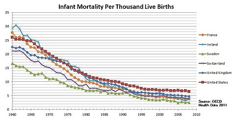
There has been a lot of speculation, lately, about vaccines possibly being harmful and, in particular, causing autism. You know I work on HIV vaccine design, so there's no need to say where I stand on the need of vaccinations. No link has been found between the incidence of autism and vaccination. Of course, medicine is not an exact science. Outliers will always exist. The U.S. seem to be a special case, as the vaccination schedule in this country requires a high number of vaccine doses, yet the infant mortality rate is one of the highest among North America and European countries. However, take a close look at this graph:


The countries with low mortality rate shown in this graph have a strict vaccination schedule, just like the U.S. On the other hand, what distinguishes them from the US is affordable health care. Countries with a high infant mortality rate are countries where poor people do not have access to vaccines and good health care. For the 3-million AIDS orphans living in sub-Saharan Africa a vaccine against HIV is the only hope they have to live into adulthood. It is quite easy for those of us who have a healthy life style and have access to food, medicines, and doctors on a daily basis, to say "no, thank you" to vaccines. But please, when you make your own decision about vaccines, do remember the millions of people for whom this is not a choice. And also remember: some children who are immunodefecient really cannot be vaccinated. They cannot contract any kind of disease, either, because their immune system is not working. However, if the majority of the people continue to get vaccinated, people who really cannot be vaccinated are still protected:

found on Facebook

Back to autism. As you saw from my last post, ASD, or autism spectrum disorders, is indeed a puzzling disease and pinning down its etiology has been challenging. The genetics involve numerous genes and diverse pathways, implying that different mechanisms could potentially lead to ASD, particularly during fetal development. One thing that I recently discovered is a number of correlations found between infections in the mother during gestation and autism:
"Recent studies have highlighted a connection between infection during pregnancy and the increased risk of autism in the offspring. Parallel studies of cerebral spinal fluid, blood and postmortem brains reveal an ongoing, hyper-responsive inflammatory-like state in many young as well as adult autism subjects. There are also indications of gastrointestinal problems in at least a subset of autistic children [1]."
In his review [1], Patterson makes a good summary of the relevant studies: for example, a permanent, inflammatory-like state has been found in postmortem examination of ASD affected brains. This was found at all ages, indicating that the state was established early in the development and maintained throughout the life-span of the ASD affected individual. These abnormalities expand to the central nervous system and the peripheral immune system affecting also the gastrointestinal tract:
"These findings include immune cell infiltrates present in the colon, ileum and duodenum, as well as increased T cell activation in the intestinal mucosa. These inflammatory changes are associated with autoimmune responses that could contribute to the observations of decreased mucosal integrity, or 'leaky gut' [1]."
"Abnormal activation of the immune system may also be involved in the etiology of autism. [. . .] Family members of autistic children, particularly the mothers, show a higher incidence of allergy or autoimmune diseases. Consistent with immune involvement are findings that maternal infection is a risk factor for autism [2]."
In conclusion, there is a correlation between immune abnormalities and ASD, and the immune abnormalities propagate to the brain and the gastrointestinal tract. However, it is unclear if these abnormalities cause the behavioral symptoms of ASD or if they are a secondary effect. The health and well-being of our immune system has such deep, profound effects on the central nervous system. The two interact very closely together: stress and the general emotional status, for example, can affect immunity; vice versa, the immune system can influence behavior. Both our brain and our immune system constantly learn and readapt to the surrounding environment (for example, our immune system learns to recognize new pathogens throughout our lifetime), which makes them prone to life-long epigenetic changes induced by environmental factors such as stress and disease. It's not a coincidence that:
"Immune dysregulation has also been implicated in the etiology of a variety of neurodegenerative, psychiatric, and neurodevelopmental disorders, including Parkinson, Huntington, and Alzheimer diseases, multiple sclerosis, major depression, schizophrenia, and addiction [2]."
Hsiao et al. [2] addressed the open question of whether the immunological abnormalities cause ASD-like behaviors in a mouse model. They induced ASD in mouse offspring through "maternal immune activation" (MIA): the immune system of pregnant mice was altered and then the offsprings of the altered mice that were behaviorally abnormal was compared to the offsprings of the controls. The behaviorally abnormal MIA offsprings exhibited core behavioral symptoms of autism, including increased repetitive behaviors, decreased social interactions, and increased anxiety. Hsiao et al. found several abnormalities in the immune system of these MIA offsprings: levels of regulatory T-cells were decreased and CD4+ T-cells were hyper-responsive. These abnormalities could not be transferred to healthy mice through a bone marrow from the MIA mice. However, when irradiated and transplanted with immunologically normal bone marrow, many of the behavioral abnormalities stopped. This would suggest that the immunological dysregulation causes the ASD-like behaviors.
"It is striking that in a mouse model of an autism environmental risk factor that exhibits the cardinal behavioral and neuropathological symptoms of autism, there is also permanent peripheral immune dysregulation. This finding provides the opportunity to explore molecular mechanisms underlying the relationship between brain dysfunction and altered immunity in the manifestation of abnormal behavior. Furthermore, this finding provides a platform for investigating how prenatal challenges can program long-term postnatal immunity, health, and disease. Maternal insult-mediated epigenetic modification in HSC and progenitor cells is one possible mechanism for how effects may be established by transient environmental changes yet persist permanently into adulthood. However, the BM transplant results suggest that the peripheral environment of the MIA offspring is also critical for maintaining a permanently modified immune state [2]."
We will never be able to prove or disprove a direct causal relation between vaccines and autism: if a child develops ASD after vaccination, unfortunately, we cannot rewind time and see if the same child, without the vaccine, would've never developed ASD in his/her lifetime. ASD typically develops in infancy, which is when the bulk of vaccines are administered. The risk of ASD is much higher (see last week's post) if there's already a family member with ASD, siblings in particular. And given the deep, complex interactions and reciprocal influence between the nervous system and the immune system it is quite possible that a sudden change in the immune system could cause some level of disruption in the nervous system. However, if the immune system is primed to such risk, a virus or any other pathogen, which cause changes in the immune system just like a vaccine does, could also cause similar disruptions. On the other hand, vaccines can potentially prevent infections that, according to these studies, do increase the risk of ASD in the baby during the first trimester of gestation.

So, as always: Read the literature, talk to your doctor, possibly to more than one, consider your family's medical history, and, whatever decision you make, make sure it is an informed decision.

[1] Patterson, PH (2011). Maternal infection and immune involvement in autism Trends in Molecular Medicine DOI: 10.1016/j.molmed.2011.03.001

[2] Hsiao EY, McBride SW, Chow J, Mazmanian SK, & Patterson PH (2012). Modeling an autism risk factor in mice leads to permanent immune dysregulation. Proceedings of the National Academy of Sciences of the United States of America, 109 (31), 12776-81 PMID: 22802640

ResearchBlogging.org

Sunday, December 8, 2013

Autism: not one disease but a spectrum of disorders; not one gene but a network of gene coexpressions.


"Autism spectrum disorder (ASD) is a lifelong developmental condition that affects about 1 in 110 individuals, with onset before the age of three years. It is characterized by abnormalities in communication, impaired social function, repetitive behaviors and restricted interests [1]."
ASD is more common among males than females, with a 4:1 male to female ratio. Numerous studies in the literature have shown evidence for a strong genetic component of autism, with a risk up to 25 times higher among siblings compared to the general population. However, if you look at the literature, you find that these numbers change pretty dramatically from study to study. This is often the case when you look at rare disorders in conjunction with rare mutations (WARNING: the rest of the paragraph is a statistical digression, feel free to skip to the next section). The smaller the effect you are trying to measure, the more subjects you will need in your study. This is also true if you are testing many variants, as for example in GWAS studies, which investigate variants in the whole genome. If the effect is big enough, you will find statistical support for your association, however, if your sample size is not big enough, the effect you are trying to measure will vary greatly from study to study. This is because the smaller the sample size, the larger the variance, which is stat jargon to say that whatever you are trying to measure (typically an increase in risk) is likely to be different if you repeat the study.

What do we know about the genetic etiology of ASD? About 10% of people diagnosed with ASD have some underlying genetic syndrome (including mitochondrial genes). About 5% are due to rare chromosome rearrangements, for example changes in the size, shape, or number of some chromosomes. Another 5% has been associated to both inherited and de novo "copy number variations" (CNV), the presence of extra copies of some genes [1]. CNV is not rare among humans, as it accounts for approximately 0.4% of the variation between unrelated genomes. Identical twins also differ in CNV, and, even though they have identical genomes, the copy number of the genes may differ between the two. Despite this, in some families with a history of ASD the proportion of de novo CNV's has been found to be up to five times higher than in families without a history of ASD. Finally, thanks to recent advances in sequencing technology, de novo point mutations throughout hundreds of genes have been found and implicated in about 15% of ASD cases [2].

In light of the variety of mutations, genes, and phenotypes associated with ASD, two studies published in the last issue of Cell addressed the following question:
"do these genetic loci converge on specific biological processes, and where does the phenotypic specificity of ASD arise, given its genetic overlap with intellectual disability (ID)? [2]"
"if and when, in what brain regions, and in which cell types specific groups of ASD-related mutations converge during human brain development [3]" ?
Of the two papers, I've so far only read the one by Willsey et al. [3], who combined their own data with already published data and identified 144 de novo "loss-of-function (LoF)" mutations, in other words, mutations that impair the functionality of the gene (hence the corresponding protein is no longer produced). They called genes with 2 or more de novo LoF mutations "hcASD", or "high confidence" ASD because statistically they had a high probability of being truly associated with ASD. They also analyzed a less-likely set of genes with only one de novo LoF mutation, which they called "pASD genes".

Next, the researchers investigated when and where these genes are expressed during brain development. The way they did this is a bit technical, but to think about it in simple terms think of it this way: (1) they needed samples from brain tissues taken at different developmental stages; (2) they needed to look not just at one gene, but at families of genes that are likely to interact together and influence one another's likelihood of getting turned "on" and "off". When a gene is turned "on", the gene is coding a protein, and we say that the gene is "expressed."

To carry on their analysis, Willsey et al. used data published by Kang et al. (Nature, 2011) from "57 clinically unremarkable postmortem brains of diverse ancestry (31 males, 26 females) that span 15 consecutive periods of neurodevelopment and adulthood from 5.7 postconceptual weeks (PCW) to 82 years." The gene expression values were determined for each gene by brain region and by postmortem brain sample. Brain regions were grouped according to transcriptional similarity during fetal development. These data were used to generate 52 gene coexpression networks, each network composed of the hcASD genes and their top correlated genes. This coexpression network analysis is a technique that's been extensively used lately to analyze patterns of co-expressions of genes. Each gene in the network is represented by a node, and any two nodes (genes) at any given time are connected if the genes are expressed at that time.

Using this set-up, the researchers were able to link the ASD genes to particular brain regions and developmental phases.
"Our analysis identifies robust, statistically significant evidence for convergence of the input set of hcASD and pASD risk genes in glutamatergic projection neurons in layers 5 and 6 of human midfetal prefrontal and primary motor-somatosensory cortex (PFC-MSC). Given the extensive genetic and phenotypic heterogeneity underlying ASD and the small fraction of risk genes that we have examined in this study, this likely represents only one of several such points of convergence. Nonetheless, the analytic approach presented here clarifies key variables relevant for productive functional studies of specific ASD genes carrying LoF mutations, providing an important step in moving from gene discovery to an actionable understanding of ASD biology [3]."
Cortical glutamatergic projection neurons (CPNs) are a class of neocortical neurons. They are called "projection" neurons because they transmit information from the neocortex to other neocortical and central nervous system regions. During development, projection neurons are generated in the neocortical germinal zone and migrate radially to their final neocortical position. In their study, Wyllsey et al found that the development of midfetal CPNs is particularly vulnerable to ASD. Furthermore, the set of ASD genes they identified as associated to ASD are functionally diverse and encode proteins found in distinct cell compartments, confirming the theory that ASD can be caused by different and distinct pathways.
"Given recent studies suggesting that as many as 1,000 genes or more could contribute to ASD (He et al., 2013; Iossifov et al., 2012; Sanders et al., 2012), our analysis has uncovered a surprising degree of developmental convergence. Despite starting with only nine hcASD seed genes, we have identified highly significant and robust evidence for the contribution of coexpression networks relevant to L5 and L6 CPNs in two overlapping periods of midfetal human development (3–5 and 4–6) corresponding to 10–24 PCW [3]."
The importance of these studies lies in the understanding of not just the genetic association per se, but in the mechanisms that drive these associations, and, most importantly, how the numerous genes interact and when.

[1] Devlin and Schrer (2012). Genetic architecture in autism spectrum disorder Genetics & Development DOI: 10.1016/j.gde.2012.03.002

[2] Neelroop N. Parikshak, Rui Luo, Alice Zhang, Hyejung Won, Jennifer K. Lowe, Vijayendran Chandran, Steve Horvath, Daniel H. Geschwind (2013). Integrative Functional Genomic Analyses Implicate Specific Molecular Pathways and Circuits in Autism Cell DOI: 10.1016/j.cell.2013.10.031

[3] A. Jeremy Willsey, Stephan J. Sanders, Mingfeng Li, Shan Dong, Andrew T. Tebbenkamp, Rebecca A. Muhle, Steven K. Reilly, Leon Lin, Sofia Fertuzinhos, Jeremy A. Miller, Michael T. Murtha, Candace Bichsel, Wei Niu, Justin Cotney, A. Gulhan Ercan-Sencicek, J (2013). Coexpression Networks Implicate Human Midfetal Deep Cortical Projection Neurons in the Pathogenesis of Autism Cell DOI: 10.1016/j.cell.2013.10.020

ResearchBlogging.org

Thursday, August 16, 2012

What's that gene for, again?


I'm always skeptical when you hear prepositions such as "gene X has function Y," as often there are very complicated mechanisms nestled between the "gene" and the "function/phenotype." If you've been following me over the past year (yes, I've been blogging for a year already, time flies!), we've learned that between-gene interactions (epistasis), and changes in gene expression (epigenetics) can completely change the picture.

Recent reviews on the use of RNA interference have given me additional reasons to be skeptical.

Gene function in vivo has been studied through a procedure called "gene knockdown," which uses RNA interference (RNAi) to "tune down" the expression of the gene. RNAi has also been used in to mimic human genetic diseases that would otherwise have no somatic equivalent in the animal world, in particular in studies aimed at discovering novel drug targets. By introducing synthetic RNA into the cell, researchers can effectively silence target genes and thus identify their functions within specific cellular processes.

It certainly is a brilliant tool, but there are several issues one needs to keep in mind when using RNAi. The target specificity, for example, is not always perfect, and several off-target effects (down-regulation of genes different from the target ones) have been documented. When this happens, you can no longer be sure of what genes, if not all, caused the observed change in phenotype. Ideally, in order to minimize off-target effects, one should repeat the experiment with different types of RNAi targeting the same gene. Rescuing the loss of function by re-inserting the mRNA (or making it "immune" to the RNAi) would also provide further evidence. However, this is very hard to realize in practice.

It gets more complicated.

Many genes regulate cellular fitness. The change observed change in phenotype, rather than reflect the knockdown gene, could instead be a direct consequence of lower cell proliferation. In addition, we tend to simplify things thinking that the relationship between gene and phenotype is linear, or that the effect from different genes is additive, when in fact such simple mathematical frameworks often don't capture the reality of the biological world. Interactions and non-linearity are difficult to model. Experiments that target multiple genes rank the results in terms of dose-responses, though such results are often contaminated by false positives and knockdown efficiencies.

All this not to say that this is the end of RNAi experiments, rather, that additional thought has to be given when interpreting the results. We still have a long way to go before we can fully encompass the complexity of our genome, and we are taking one baby step at the time.

William G. Kaelin Jr. (2012). Use and Abuse of RNAi to Study Mammalian Gene Function Science, 337 (6093), 421-422 DOI: 10.1126/science.1225787

ResearchBlogging.org


Monday, March 5, 2012

It takes all the running you can do to keep in the same place

"Now, here, you see, it takes all the running you can do, to keep in the same place. If you want to get somewhere else, you must run at least twice as fast as that!"
~Lewis Carroll, Through the Looking Glass
The Red Queen Effect is a genetic effect named after Lewis Carroll's famous quote, "It takes all the running you can do to keep in the same place." As it turns out, Carroll's witty and paradoxical thinking fits viral host evolution: out of the whole viral population, only a few manage to infect new hosts -- these are the viruses that are fit enough to overcome the first hurdle (bottleneck) of "jumping" into a new host. Once there, though, these viral particles are not necessarily fit to survive the new host's environment because the defenses they have developed have been selected for in the previous host (this in genetics is called "fitness cost"). Therefore, they must mutate rapidly in order to acquire new defenses that will allow them to escape the new immune system.

Do you see why Carroll's Red Queen applies? A virus has to do all the running (mutating) it takes in order to keep (survive) in the same place (host).

Variety in hosts' immune responses is ensured by the MHC genes, the major histocompatibility complex genes in vertebrates. These genes, which are the most polymorphic among vertebrates, encode molecules found on all cell surfaces that mediate antigen presentation: when an antigen (any object, either a molecule or another organism, that is recognized as a "non-self" by the body) enters a cell, it is broken up and the bits of proteins are transported to the cell surface for "presentation" to the immune system. High variability in this class of molecules ensures that a wide range of antigens can be recognized and hence trigger the immune response. The high variability found in the MHC genes is the reason for the high variation in disease susceptibility in the population, for example, why a particular flu strain can keep one person in bed for a whole week, while another only gets a mild cold for a couple of days.

Just like the virus needs to do a lot of running in order to overcome the immune system, the immune system itself is at an advantage the more antigens it is able to recognize. So, you see, the Red Queen Effect, applies to both the host and the pathogen, leading to an antagonistic coevolution that ensures diversity in the MHC genes in the population.

A recent PNAS paper [1] investigates how this mechanism is maintained:
"One leading explanation, antagonistic coevolution (also known as the Red Queen), postulates a never-ending molecular arms race where pathogens evolve to evade immune recognition by common MHC alleles, which in turn provides a selective advantage to hosts carrying rare MHC alleles. This cyclical process leads to negative frequency-dependent selection and promotes MHC diversity if two conditions are met: (i) pathogen adaptation must produce trade-offs that result in pathogen fitness being higher in familiar (i.e., host MHC genotype adapted to) vs. unfamiliar host MHC genotypes; and (ii) this adaptation must produce correlated patterns of virulence (i.e., disease severity)."
In [1], Kubinak et al. describe how they repeatedly transmitted the same pathogen through different hosts (groups of genetically identical mice, each group carrying a different MHC family) and observed patterns of pathogen adaptation.
"Results from our experiments demonstrate that pathogen adaptation is host MHC genotype-specific. We conclude that pathogen adaptation to the familiar host MHC genotype produces trade-offs in pathogen fitness by reducing the reproductive output of adapted viruses when infecting hosts carrying unfamiliar MHC genotypes. Although previous work has shown that interactions between host and pathogen genotypes are important for determining patterns of pathogen fitness and virulence associated with infection, our dataset is unique in that it provides direct experimental support for fitness trade-offs associated with a pathogen’s adaptation to specific host MHC genotypes, thus confirming the first major assumption of the antagonistic coevolution model of MHC evolution."
With their experiment, Kubinak and colleagues proved that diversity in the MCH gene complex is maintained through the antagonistic coevolution between pathogen and host. This does not exclude other factors such as the heterozygote advantage and mating preferences, which have also been considered as likely explanations of the high variability in this gene family. As it often happens in genetics, the likely explanation is an interaction between all these phenomena and the hardest thing is to disentangle each contribution.

The authors conclude with one final thought, as their results suggest that populations with low MHC genetic diversity are likely to select for more virulent pathogens.
"If this suggestion is true, many livestock breeds and endangered species that exhibit reduced genetic diversity may be particularly sensitive to the consequences of rapid pathogen adaptation. Additionally, the prophylactic use of antibiotics on domesticated livestock places a selective pressure on pathogen populations to evolve antibiotic resistance, and as a consequence has been implicated in the emergence of antibiotic-resistant strains of human pathogens."

[1] Kubinak, J., Ruff, J., Hyzer, C., Slev, P., & Potts, W. (2012). From the Cover: Experimental viral evolution to specific host MHC genotypes reveals fitness and virulence trade-offs in alternative MHC types Proceedings of the National Academy of Sciences, 109 (9), 3422-3427 DOI: 10.1073/pnas.1112633109

ResearchBlogging.org