Debunking myths on genetics and DNA

Showing posts with label genes. Show all posts

Friday, April 29, 2016

Hunting For The Signatures of Cancer

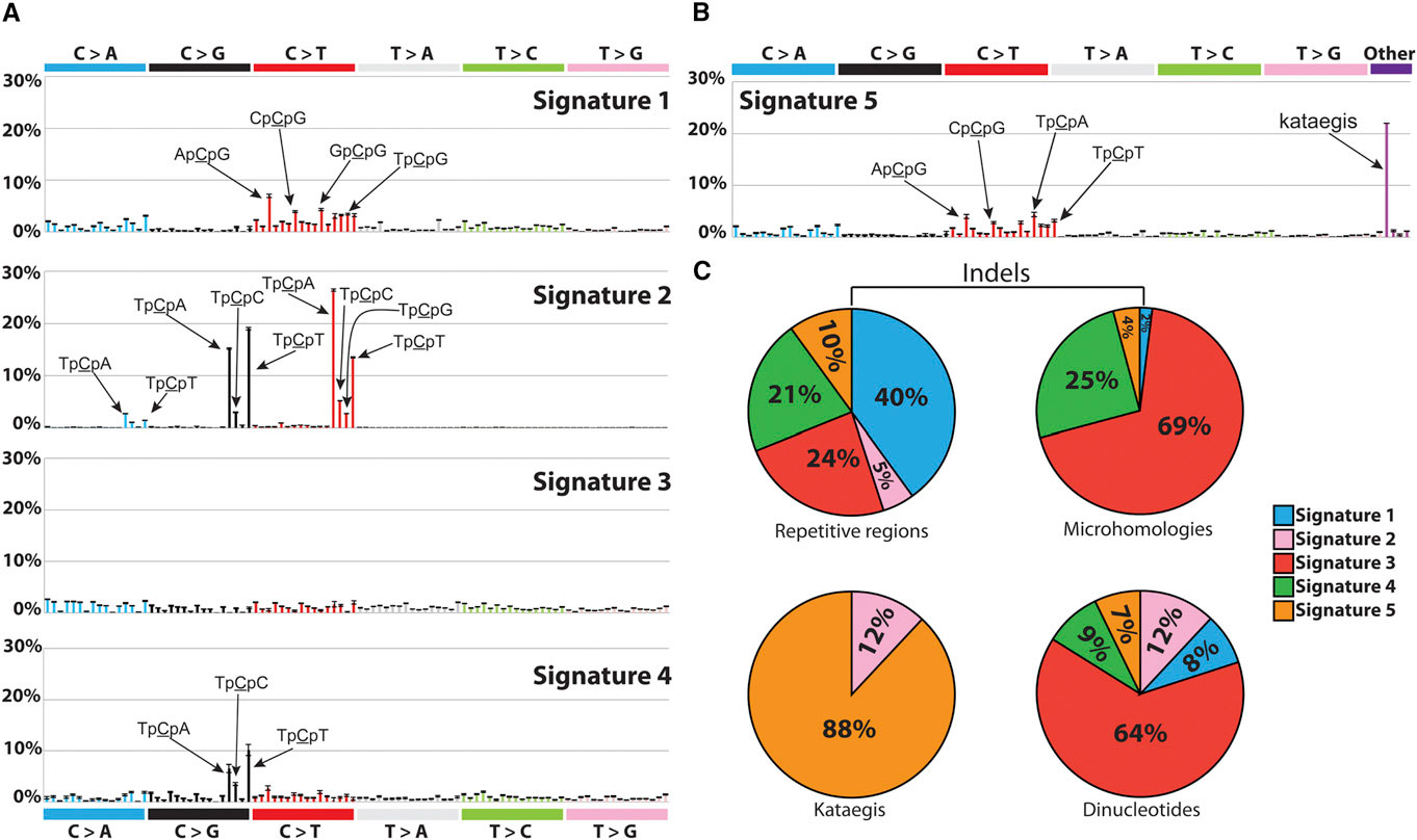
Signatures of Mutational Processes Extracted from the Mutational Catalogs of 21 Breast Cancer Genomes. Credit:http://dx.doi.org/10.1016/j.celrep.2012.12.008

Cancer is the second leading cause of death worldwide, with approximately 14 million new cases and 8.2 million cancer related deaths each year (Source: WHO). A family history of cancer typically increases a person's risk of developing the disease, yet most cancer cases have no family history at all. This suggests that a combination of both genetics and environmental exposures contribute to the etiology of cancer. In this context, "genetics" means the genetic make-up we are born with and inherited from our parents. For example, women born with specific mutations in the BRCA1 and BRCA2 genes are known to have a much higher risk of developing breast cancer later in life.

However, besides the genetic make-up we carry from birth, there are many geographical and environmental factors that contribute to the risk of cancer. For example, the incidence of breast cancer is over 4 times higher in North and West Europe compared to Asia and Africa (Source: WHO). Stomach cancer, on the other hand, is much more prevalent in Asia than the US. If you think that this may be linked to the genetic differences across ethnicities, think again. The National Cancer Institute published a summary of several studies that compared the incidence of first and second generation immigrants in the US with the local population. They found that:
"cancer incidence patterns among first-generation immigrants were nearly identical to those of their native country, but through subsequent generations, these patterns evolved to resemble those found in the United States. This was true especially for cancers related to hormones, such as breast, prostate, and ovarian cancer and neoplasms of the uterine corpus and cancers attributable to westernized diets, such as colorectal malignancies."
According to the World Health Organization (WHO),
"around one third of cancer deaths are due to the 5 leading behavioral and dietary risks: high body mass index, low fruit and vegetable intake, lack of physical activity, tobacco use, alcohol use."
Cancer is the result of a series of cellular mechanisms gone awry: every time a cell divides, somatic mutations accumulate in the cell's genome. These are not mutations we are born with, inherited from our parents. Rather, these are changes that accumulate in certain cells as we grow old and are not  the same across all cells in the body. Many environmental exposures contribute to this process and affect the rate at which these mutations accumulate. However, cells have various mechanisms that are normally able to repair harmful mutations or, when the damage is beyond repair, to trigger cell death. The immune system is also "trained" to recognize cancer cells and destroy them.

When all these defense mechanisms fail, cancer cells start dividing uncontrollably.

As a result, all cancer cells carry a number of somatic mutations that set them apart from healthy cells, and some tend to be the same across different cancer patients: for example, specific mutational patterns found in lung cancer have been attributed to tobacco exposure and were indeed reproduced in animal models. Another set of mutations has been attributed to UV exposure and has been found in skin cancers [1, 2].

This prompts the ambitious question: can we find common mutations across individuals with the same cancer? And how many of these mutational patterns that are common across individuals can we attribute to particular exposures and/or biological processes? Distinguished postdoctoral researcher Ludmil Alexandrov, from the Los Alamos National Laboratory, has been working on this problem since his he was a PhD student at the Wellcome Trust Sanger Institute.

"It's like lifting fingerprints," Alexandrov explains. "The mutations are the fingerprints, but now we have to do the investigative work and find the 'perpetrator', i.e., the carcinogens that caused them." During his graduate studies, under the supervision of Mike Stratton of the Wellcome Trust Sanger Institute, Alexandrov developed a mathematical model that, given the cancer genomes from a number of patients, is able to pick the "common signals" across the patients -- i.e. mutation patterns that are common across the patients -- and classify them into "signatures."

"When formulated mathematically," Alexandrov explains, "the question can be expressed as the classic 'cocktail party' problem, where multiple people in a room are speaking simultaneously while several microphones placed at different locations are recording the conversations. Each microphone captures a combination of all sounds and the problem is to identify the individual conversations from all the recordings." Taking from this analogy, each cancer genome is a "recording", and the task of the mathematical model is to reconstruct each conversation, in other words, the mutational patterns. These are sets of somatic mutations that are the observed across the cancer genomes and that characterize certain types of cancers.

In 2013, Alexandrov and colleagues analyzed 4,938,362 mutations from 7,042 patients, spanning 30 different cancers, and extracted more than 20 distinct mutational signatures [2]. "Some patterns were expected, like the known ones caused by tobacco and UV light," Alexandrov says. "Others were completely new."

Of the new signatures found, many are involved in defective DNA repair mechanisms, suggesting that drugs targeting these specific mechanisms may benefit cancers exhibiting these signatures [3]. But the most exciting part of this research will be finding the 'perpetrator' or, as Alexandrov explains, the mutations triggered by carcinogens like tobacco, UV radiation, obesity, and so on. The challenge will be to experimentally associate these mutational patterns to the exposures that caused them. In order to do this, the scientists will have to expose cultured cells and model organisms to known carcinogens and then analyze the genomes of the experimentally induced cancers.

In the meantime, the signatures found so far are only the beginning: Alexandrov and colleagues have teamed up with the Los Alamos High Performance Computing Organization in order to analyze the genomes of almost 30,000 cancer patients.

"The amount of data we will have to handle for this task is enormous, on the order of petabytes," Alexandrov says. "Few places in the world have the capability to handle this many data. Under normal circumstances, it takes months to answer a question on 10 petabytes of data. The supercomputing facility at Los Alamos can provide an answer within a day."

Because of his research, in 2014 Alexandrov was listed by Forbes magazine as one of the “30 brightest stars under the age of 30” in the field of Science and Healthcare. In 2015 he was awarded the AAAS Science & SciLifeLab Prize for Young Scientists in the category Genomics and Proteomics [2] and the Weintraub Award for Graduate Research. He is now the recipient of the prestigious Oppenheimer fellowship at Los Alamos National Laboratory.

References
Siegel, R., Miller, K., & Jemal, A. (2015). Cancer statistics, 2015 CA: A Cancer Journal for Clinicians, 65 (1), 5-29 DOI: 10.3322/caac.21254

[1] Alexandrov LB (2015). Understanding the origins of human cancer. Science (New York, N.Y.), 350 (6265) PMID: 26785464

[2] Alexandrov LB, Nik-Zainal S, Wedge DC, Aparicio SA, Behjati S, Biankin AV, Bignell GR, Bolli N, Borg A, Børresen-Dale AL, Boyault S, Burkhardt B, Butler AP, Caldas C, Davies HR, Desmedt C, Eils R, Eyfjörd JE, Foekens JA, Greaves M, Hosoda F, Hutter B, Ilicic T, Imbeaud S, Imielinski M, Jäger N, Jones DT, Jones D, Knappskog S, Kool M, Lakhani SR, López-Otín C, Martin S, Munshi NC, Nakamura H, Northcott PA, Pajic M, Papaemmanuil E, Paradiso A, Pearson JV, Puente XS, Raine K, Ramakrishna M, Richardson AL, Richter J, Rosenstiel P, Schlesner M, Schumacher TN, Span PN, Teague JW, Totoki Y, Tutt AN, Valdés-Mas R, van Buuren MM, van 't Veer L, Vincent-Salomon A, Waddell N, Yates LR, Australian Pancreatic Cancer Genome Initiative, ICGC Breast Cancer Consortium, ICGC MMML-Seq Consortium, ICGC PedBrain, Zucman-Rossi J, Futreal PA, McDermott U, Lichter P, Meyerson M, Grimmond SM, Siebert R, Campo E, Shibata T, Pfister SM, Campbell PJ, & Stratton MR (2013). Signatures of mutational processes in human cancer. Nature, 500 (7463), 415-21 PMID: 23945592

[3] Alexandrov LB, Nik-Zainal S, Siu HC, Leung SY, & Stratton MR (2015). A mutational signature in gastric cancer suggests therapeutic strategies. Nature communications, 6 PMID: 26511885

ResearchBlogging.org

Wednesday, March 16, 2016

An open letter to all science lovers who want to defend science ... please don't.


Last week I had an animated discussion on Facebook over an older post in which I describe some literature I dug out on possible (underline “possible”!) correlations with autism. True, my post is highly incomplete, but it was meant as a discussion starter to point at things that scientists have been looking at in an attempt to unravel what feels like a rise in autism. Is autism the new childhood plague of our modern society or has it always been around and we just became more aware of it? And if the rise is real, what caused it?

To me the most intriguing bit is that if you type 'autism gut microbiota' into the PubMed search field (for those not familiar with PubMed, it's a repository for medical literature), you find an incredible number of studies and reviews: apparently there is an association between autism and disruptions of the gut microbiota, but whether the two are truly correlated or the correlation is spurious is still unclear.

Before I go on analyzing the literature I found on this topic, let me open a parenthesis on the Facebook discussion because it's something I deeply care about. You might think that the animated discussion I got into was with anti-vaxxers who believe that vaccines cause autism. Instead, my post was criticized by pro-vaccine people who, with the same unflinching certainty typical of the anti-vaxxers, believe that the rise in autism is fiction invented by anti-vaxxers, that autism has always been around, and that any difference between gut microbiota of autistic children and non-autistic children has been disproved. "By whom?" I asked. By this one report:
"Children with autism have no unique pattern of abnormal results on endoscopy or other tests for gastrointestinal (GI) disorders, compared to non-autistic children with GI symptoms, reports a study in the Journal of Pediatric Gastroenterology and Nutrition."
Notice that this opening line is a bit misleading because here is the actual paper [1] whose conclusion, quoting from the abstract, are a bit more cautiously stated:
"This study supports the observation that children with autism who have symptoms of gastrointestinal disorders have objective findings similar to children without autism. Neither non-invasive testing nor endoscopic findings identify gastrointestinal pathology specific to autism, but may be of benefit in identifying children with autism who have atypical symptoms."
Notice also the difference from the abstract and the title of the report. You can tell which one was written by a scientist, right? Because when you do a search on PubMed using keywords autism and gut microbiota you find a long list of references and decades of research. So to me what this says is that the question is still open and we need to understand the issues better. It takes way more than one paper to disprove hypothesis-raising questions spurred from decades of research.

Now here's the mother of all problems: the Internet has made everyone (EVERYONE!) an expert. Today you no longer need a medical degree to speak authoritatively about vaccines, disease, and health. This has generated movements like the anti-vaxxers, but, even more unfortunate is the rise of groups that reply to the anti-vaxxers without a scientific mind-set: these people are doing even more damage to the community than the anti-vaxxers themselves. I found myself in a conversation that had the same one-ended arguments used by anti-vaxxers except these were people who are actually in favor of vaccines: for every paper on autism and gut microbiota I brought up they would dismiss it with another one that said the opposite, demonstrating no understanding of the difference between raising hypotheses and making a claim.

As a scientist, I can tell you that this behavior is the very opposite of scientific thinking. All the people who are in favor of science but DO NOT adopt a scientific attitude when counter-arguing non-scientific claims are hurting the scientific community. It's happening for vaccines, for evolution, and for global warming. For example, people who support intelligent design are mistaken about evolution because they don't understand the meaning of the word "theory" and they don't understand how scientific thinking works. We need to educate people on scientific thinking, not give bad examples of undebatable and absolute notions.

So, PLEASE, all science fans, I beg of you: support us by giving us a cheer, by always citing original papers, and by keeping an open mind because that's what a real scientist would do. We are raising hypotheses, not discussing the meaning of Bible verses. And if you know you can't do any of the above, then the best support you can give us is to shut up. Let real science speak for itself.

I'm fully aware that I'm preaching to the choir so I'll stop now and resume my discussion on autism and gut microbiota. As an additional side note, let me emphasize how difficult it is to discuss a topic like autism because of its extreme complexity: it's a relatively new diagnosis (first described in the early twentieth century), and even though no exact etiology has been found of date, the genetic studies conducted so far have implicated as many as 400 genes such that a malfunction in any of these genes could possibly result in autism [2].

Let's start from the facts: our body hosts more microbial cells than human cells, with the vast majority residing in the gut. These organisms, which we collectively call the "human microbiota" (and “gut microbiota” when referring to the ones residing in the gut) interact with our cells in symbiosis and in fact, some experiments have shown that they can affect our health and even gene expression (see this old post for a striking example of how genes expressed by gut bacteria can affect whether we are fat or lean). All this has been known for a long time, but it's only recently that, thanks to the advent of new DNA sequencing techniques that scientists have been able to look deeper into the composition and classification of the human microbiota. Metagenomic studies have found over 3 million distinct microbial genes (collectively called the "microbiome") in human stools, which is astonishing if you think that the human genome, in comparison, contains about 20-30 thousand genes. The gut microbiome is rich in enzymes without which our body would be unable to digest important nutrients. In fact, it's estimated that roughly 10% of our dietary energy intake comes from byproducts of fermentation from the gut bacteria.

That's all fine and dandy, but what does this have to do with behavior and brain health? A lot, actually, to the point that scientists coined the phrase "gut-brain axis" to denote the deep interaction between the nervous system and the gut microbiota. A 2011 PNAS study [3] used a mouse model to demonstrate how the gut microbiota affects mammalian brain development and behavior. This can happen in a number of ways, but one interesting hypothesis is that a healthy gut microbiome can help modulate the concentration of chemicals that are important for brain development as well as important nutrients that are precursors of neurotransmitters like serotonin.

Several studies done on different populations of children affected by autism spectrum disorders (ASD) have reported some form of gastro-intestinal (GI) dysfunction (such as food intolerances, abdominal pain, diarrhea and flatulence), with proportions ranging from 20-60% of the study population [4]. It's true that ASD children are often very picky eaters with drastic dietary habits, which would of course cause the GI issues. However, given the previously mentioned evidence that the gut microbiota shapes brain development since early infancy, the question of which is the cause and which is the effect at this point is legitimate. In other words, what came first, the chicken or the egg?

Studies have pointed at alterations of the gut microbiota in ASD children who experience gastro-intestinal issues, and some have reported that ASD children receiving antibiotics seemed to experience behavioral improvements. Drastic changes in diet (for example adopting a gluten-free and/or casein free diet) have shown behavioral improvements in some ASD studies, but not in all (meaning that some studies still didn't observe any improvement). Some papers report a higher risk of ASD in children who have not been breast-fed or who have been weaned after the first month of life. All of these instances would cause the gut microbiota to change, including breast feeding, which plays a fundamental role in establishing a healthy bacterial flora in infants. But why aren't any of these studies conclusive? And why are some conclusions the opposite of others? Such differences in results can be explained by differences in sample sizes (too few patients, for example, would cause a false negative), and also by the fact that many of these children have impaired communication skills, and therefore the symptoms, rather than being self-reported, are gathered from the observations of the parents, which can potentially introduce a bias.

Studies that have compared the microbial composition of stools in children affected by ASD with healthy children have had mixed results: the majority report some differences in the composition of the microbial populations, while a few found no significant differences. And despite many studies have looked into it, no ASD-specific gut disturbance has been found, meaning that whatever gut issues ASD children may experience, they are no different than the ones healthy children may experience as well. At the same time, there is some evidence that probiotics help relieve some of the gastro-intestinal issues ASD children experience and at the same time, improve some of their behavioral issues.

What conclusion can we draw from this? Well, first of all that there's no black and white but a lot of gray and anyone who will tell you it's either black or white does not understand how science works. Look at Lamarck's theory of the evolution of traits, first dismissed by Darwin and now (sort of) coming back in the form of epigenetics. Science is not a means to get a definitive and absolute truth, rather, it is our drive to keep asking questions in the search for working answers. [On a side note, this is exactly why I do not like certain showmen out there who proclaim themselves scientists just because they promote science "truths"; real science educators should be promoting scientific thinking, instead.] More than once in the history of science we've corrected and generalized theories. That doesn't mean that we were wrong, rather, it means that we've expanded our knowledge and acquired better investigative tools.

Unfortunately we don't have historic data on autism, since the term was first used in the early 1900s and the definition of the disorder has changed over time. This questions whether or not case prevalence has been truly rising over time, or, instead, the rise we're seeing is simply the effect of a more comprehensive diagnosis. Regardless of whether this is true or not, the fact that most cases are reported in industrialized countries raises an important speculation: these are countries that have seen the most drastic dietary changes over the past 100 years and also lifestyle changes in terms of hygiene and use of antibacterial products, both in household items, as well as in livestock farming (and the use of antibiotics in livestock farming has indeed been increasing over the past few decades). There is no denying that dietary changes and increased use in antimicrobial products will affect the bacteria coexisting in our environment. Are these changes significant? Can they be play a role in the rise in autism prevalence? Can they play a role in the etiology of other disease whose prevalence appears to be on the rise, such as asthma, food allergies, and autoimmune disorders?

I do believe that these are legitimate questions that call for a deeper understanding of how our body interacts with the environment, both outside and inside. Throughout time, evolution has provided us with ways to adapt, but such adaptations are slow. Instead, over the past 100 years we've introduced drastic changes both in the environment as well as in our lifestyle in ways that are too fast for our genetic make-up to adapt. Anything concerning humans is complex, layered by multiple interactions between genetics, environment, and behavior. That’s why we need to keep looking and, most importantly, that’s why we need to always keep an open mind on things. Anyone who claims to know the absolute truth has misunderstood what science is about. Fighting bogus facts like the ones brought forth by the anti-vaxxers with analogous “absolute truths” will only reinforce the globally spread misunderstanding of what science is and what function it covers in our path toward understanding the world. The day we stop asking questions because we’ve found all the answers is the day we’ve stopped growing.

[1] Kushak RI, Buie TM, Murray KF, Newburg DS, Chen C, Nestoridi E, & Winter HS (2016). Evaluation of Intestinal Function in Children with Autism and Gastrointestinal Symptoms. Journal of pediatric gastroenterology and nutrition PMID: 26913756

[2] Li, Q., & Zhou, J. (2016). The microbiota–gut–brain axis and its potential therapeutic role in autism spectrum disorder Neuroscience DOI: 10.1016/j.neuroscience.2016.03.013

[3] Heijtz, R., Wang, S., Anuar, F., Qian, Y., Bjorkholm, B., Samuelsson, A., Hibberd, M., Forssberg, H., & Pettersson, S. (2011). Normal gut microbiota modulates brain development and behavior Proceedings of the National Academy of Sciences, 108 (7), 3047-3052 DOI: 10.1073/pnas.1010529108

[4] Mulle, J., Sharp, W., & Cubells, J. (2013). The Gut Microbiome: A New Frontier in Autism Research Current Psychiatry Reports, 15 (2) DOI: 10.1007/s11920-012-0337-0

ResearchBlogging.org


Monday, February 15, 2016

Decoding the Dark Matter of the Human Genome

First appeared on my Huffington Post blog on February 15, 2016. 

In 1994, researchers from Harvard and Stanford published a paper in which they described three mice: one was yellow and fat, one mottled and fat, and the last one was brown and lean. An ordinary image, except for one thing: despite being so different, all three mice were genetically identical.

If their genes were exactly the same, what was causing such striking differences in the mice?

Three genetically identical mice that do not look the same. Why?
Photo credit: Nature Publishing, used with permission

At the time, Karissa Sanbonmatsu--staff scientist at Los Alamos National Laboratory--was working on plasma physics, and she had no idea that one day she would tap into this mystery. Even though she started from a completely different field, from the very beginning she was obsessed by one question: What distinguishes life from matter?

"In order to answer that question, the first place to look is the ribosome," Karissa explains. "It's the oldest molecule found in life."

And for a reason: all living cells are made of proteins, and ribosomes are the "factory" inside the cell where these proteins are made.

The breakthrough came in 2003, when the Q Machine, at the time the second fastest supercomputer in the world, was built at Los Alamos National Laboratory. Using the Q Machine, Karissa and colleagues were able to run the largest simulation ever performed until then in biology, allowing them to be the first team to publish an atomic structure of a ribosome in 2004.

This milestone set the foundation for a deeper understanding of the ribosome. Possible future applications, for example, include making new cancer therapies based on how ribosomes differentiate in healthy versus cancerous tissue.

In the meantime, a new, emerging field had been revolutionizing the way we think of genetics and inheritance: epigenetics. The three lab mice from 1994 were one example of how, by switching genes on and off, genetically identical individuals could have different observable characteristics ("phenotypes"). Epigenetics is the field that studies the mechanisms by which the environment can trigger these on/off gene patterns (called gene expression patterns), and how these modifications can be passed on to the next generation.

Both animal and human studies have shown that traits acquired by the parents, such as stress responses or the ability to store fat, can be passed on to their offspring. While DNA remains unaltered, what triggers these changes in phenotype is the activation or deactivation of genes--in other words, whether certain genes produce the proteins they code for.

But how are genes turned on or off? Specific factors regulate whether a gene is expressed (turned on) or silenced (turned off). These factors are recruited by RNA, the single-stranded molecule implicated in numerous cellular processes, from coding and decoding genes to protein synthesis.

When they were first discovered, RNA and DNA molecules that didn't code for proteins were dubbed the "dark matter" of the genome because their function was unknown. Today we know that these molecules can affect gene expression and even change traits by turning on or off certain genes.

That RNA had the power to turn genes off has been known since the early 2000s, when small RNAs were used to create mice whose cells had one particular gene silenced. Larger RNA molecules that don't code for any specific protein can also be found in different sizes inside the cell. Called long non-coding RNAs (lncRNA), they are present in great numbers in stem cells and embryos and are essential in many developmental processes.

"RNA could be the missing link in epigenetics," Karissa explains. "Ribosomes are made of RNA, and so, for me, the leap from ribosomes to lncRNAs was a natural one."

In order to understand how lncRNAs can turn genes on and off, scientists first need to unveil their molecular structure. Can lncRNAs assume different shapes, or 3D structures, and change function accordingly, or are they bidimensional molecules? Karissa and colleagues are determined to solve the puzzle. The same techniques used to resolve the ribosome structure in 2005 can be applied to lncRNAs, but because of their larger size, the team will need faster and better computational tools than the ones they used 10 years ago.

Luckily, next-generation supercomputing is underway at Los Alamos with the construction of Trinity, a machine fast enough to accommodate simulations of 3D atomic structures. This is where Karissa and colleagues are planning to run their lncRNA models.

Revealing the shape of lncRNAs would be a breakthrough. But for Karissa and her team, another even more ambitious project is on the way: "Thanks to the amazing resources offered by Trinity, we will be able to run the first atomistic simulation of human chromatin, the big 'yarn' of DNA and proteins that sits inside the cell nucleus."

Source: National Institutes of Health

This means simulating the 3D structure of three billion base pairs, plus all the proteins the DNA is wrapped around! All genes reside inside the chromatin, and this is where they are activated or deactivated. Therefore, solving the 3D structure of the chromatin will shed new light on the epigenetic mechanisms that regulate gene expression.

Many diseases are characterized by altered gene expression. For example, DNA-repairing genes are turned off in cancer cells, while genes that promote replication are over-expressed. Understanding the mechanisms that lead to these altered on/off patterns and how to reverse them can pave the way to new therapies and more efficient treatments--a bright future indeed for molecules once dismissed as the genome's dark matter.

Elena E. Giorgi is a computational biologist in the Theoretical Division (Theoretical Biology group) at the Los Alamos National Laboratory and the author of the science fiction thrillers Chimeras, Mosaics, and Gene Cards.

References
Karissa Sanbonmatsu's TEDx talk "How You Know You're in Love: Epigenetics, Stress & Gender Identity."

Duhl DM, Vrieling H, Miller KA, Wolff GL, & Barsh GS (1994). Neomorphic agouti mutations in obese yellow mice. Nature genetics, 8 (1), 59-65 PMID: 7987393

Tung CS, & Sanbonmatsu KY (2004). Atomic model of the Thermus thermophilus 70S ribosome developed in silico. Biophysical journal, 87 (4), 2714-22 PMID: 15454463

Sanbonmatsu KY, Joseph S, & Tung CS (2005). Simulating movement of tRNA into the ribosome during decoding. Proceedings of the National Academy of Sciences of the United States of America, 102 (44), 15854-9 PMID: 16249344

Structural architecture of the human long non-coding RNA, steroid receptor RNA activator. Novikova IV1, Hennelly SP, Sanbonmatsu KY. Nucleic Acids Res. 2012 Jun;40(11):5034-51. doi: 10.1093/nar/gks071. Epub 2012 Feb 22. PMID: 22362738
Sanbonmatsu KY (2016). Towards structural classification of long non-coding RNAs. Biochimica et biophysica acta, 1859 (1), 41-5 PMID: 26537437

Friday, December 18, 2015

Scientists reproduce a stress-induced phenotype in mouse pups thanks to epigenetic reprogramming

© Elena E. Giorgi

I'm excited to be blogging about science again, albeit only occasionally. Those of you who have been following the blog from its very beginnings, back in 2011, know that I've always been fascinated with epigenetics, one of my favorite topics to discuss. So much so that I've managed to include it into the plot of my detective thriller Chimeras. The thrills in the book are fictional, but the science is all real.

I was talking with my colleague Karissa Sanbonmatsu last week, who's been working on RNA and epigenetics since the early 2000s, and she was telling me how the field is still riddled with controversy. There's more and more evidence that environmentally triggered traits like stress, fat storage, and the propensity to acquire certain diseases can be passed on from one generation to the next via activated epigenetic marks, yet many scientists still refuse to believe it. How can things that are not encoded in the DNA be transmitted to the new generation? Germ cells carry epigenetic signatures that have been shaped by the environmental exposures from the parents, but how are these signatures communicated across generations?

A little background.
Our cells carry long bits of RNA that sense molecules and their changes in concentrations. Depending on the environmental exposures they find, they recruit epigenetic factors that then activate certain genes and/or deactivate others. This happens by inducing changes in the chromatin, the big yarn of DNA that sits inside the nucleus. When a gene needs to be activated, the big yarn moves until that particular gene is exposed on the surface and then translated into proteins. On the other hand, to silence the gene, the chromosome move around again and "hide" the gene deep inside the chromatin. RNA molecule act as regulators of these mechanisms, "deciding" which genes to activate and which ones to silence.

A recent study published on PNAS sheds new light on the mechanisms that communicate epigenetic marks from the germ line to the offspring, proving that epigenetic signatures acquired by the parents can be passed onto the offspring. Rodgers et al., from the University of Pennsylvania, used a mouse model to establish the following points:

  • First, they exposed male mice to chronic stress prior to breeding, and then observed reprogramming of certain genes in the hypothalamus of the offspring;
  • Second, they looked at the sperm of the stressed mice and compared it to the sperm of non-stressed mice; they found a change in content of micro RNAs (miRNAs), and 9 miRNA molecules in particular were found in much higher concentrations in the stressed mice's sperm [1]. Rodger et al. hypothesized that the 9 miRNAs were responsible for the genetic reprogramming induced by the chronic stress exposure and passed on through the paternal line.

To prove it, they injected the 9 miRNAs into single-cell zygotes that were then implanted into normal female mice, raised with no stress exposure, and then examined to see if they presented the same stress phenotype observed in the stressed male's offspring. Indeed, expression of the target genes in the hypothalamus was reduced in the mice that originated from these zygotes, and the expression patterns observed in these mice recapitulated what they had observed in the offspring of the stressed mice.

This study, published in PNAS last october [2], is a milestone in epigenetics, as it finally shows a molecular mechanism that allows genetic reprogramming in the parent to be transmitted to the offspring.

As a final thought, I want to toss in my two cents on the debated rise of autism spectrum and ADHD disorders currently observed in the Western world. Of course, there's the caveat that the diagnostic methods have changed drastically in the past few decades. Still, the increase seems real and the sad truth is that there's probably more than one cause, and the causes lie not just in what the child has been exposed to, but, once you throw in epigenetics into the pictures, his/her parents and grandparents as well. My parents for example grew up at the peak use of asbestos, DDT, and lead in paint. Yes, they survived and, knock on wood, they are quite healthy in fact. But I do fear that we will carry the consequences of those exposures for a few more generations. And who knows what the current exposure to the massive use of corn syrup and antibiotics will do to future generations. Food for thought.

[1] Rodgers AB, Morgan CP, Bronson SL, Revello S, & Bale TL (2013). Paternal stress exposure alters sperm microRNA content and reprograms offspring HPA stress axis regulation. The Journal of neuroscience : the official journal of the Society for Neuroscience, 33 (21), 9003-12 PMID: 23699511

[2] Rodgers, A., Morgan, C., Leu, N., & Bale, T. (2015). Transgenerational epigenetic programming via sperm microRNA recapitulates effects of paternal stress Proceedings of the National Academy of Sciences DOI: 10.1073/pnas.1508347112

ResearchBlogging.org

Wednesday, May 21, 2014

The Human Knock-out: looking for non-working genes

© EEG

The word "knock-out" in biology is used for lab animals like mice, for example, when one of their genes is silenced in order to study the effects of not having that gene. Silencing a gene (or knocking it out, hence the nomenclature "knock-out mouse") means that gene is no longer producing the protein it codes for. This is a condition sought for in situations where you have to test for a drug and hence the first step is to reproduce the genetic condition that caused the disease.

Mice are often "humanized", i.e. genetically engineered to carry human genes so that the experiment can be a better model for drug or therapy testing. Unfortunately, even when humanized, mice or lab animals in general are poor models for humans. When things don't work out in an animal model, we know that the experiment should not be carried out on to humans, but when on the other hand things go well in an animal experiment, there is no guarantee that it will work on humans too.

"Human chip" technology is a very promising solution, as it would bypass the need of animal testing for drug discovery. The idea is to have cell cultures from different organs on a "chip" the size of a smart phone. Lung, liver, kidney chips have already been designed and tested, but lately there has been an even further advance in making the chips part of a network connected by "blood vessels": Athena (Advanced Tissue-engineered Human Ectypal Network Analyzer) is an ongoing project to see how four organ chips (liver, lung, heart and kidney), connected by tubed filled with artificial blood, can effectively simulate a human body for drug testing and toxin screening. Athena, also dubbed the "desktop human" as given its size it would conveniently sit on a desktop, is a $19 million dollars project that will be built in the next five years.
You can read the full story here.

Athena, however, only has four organs and is still poor surrogate of the human body. The ideal solution would be to have human knock-outs to study the true effect of drugs, which of course is a little unethical to pursue. Unless human knock-outs already exist in nature. Well, guess what? They do, and they are far more common than we originally thought: on average every person has about 20 inactivated genes [1]. Wait, it gets better. Because, you may wonder, if they are so common, how come we never noticed? The ~20 inactivated genes must have some effects and/or symptoms, right?

Not necessarily. Yes, that's the most amazing thing: how robust our DNA is. People can have inactivated genes and still be healthy. It doesn't always happen, yet there are some cases when deficient gene copies are somehow compensated by other genes. And that's exactly why studying these human knock-outs is so relevant: we need to understand how people can stay healthy even when lacking important genes, as this can give new insight in drug discovery and therapy development.

In [1], MacArthur et al. screened close to 3,000 variants predicted to cause loss of gene function from 185 human genomes. Then challenge is to distinguish the "true" loss of function variants from sequencing errors. The researchers designed a "filter" to distinguish the "true" variants from the artificial errors. To me, the most striking discovery they made is that loss of function doesn't work as an "on/off" switch, rather, it can lead to a range of possible scenarios:
"Homozygous inactivation of a gene can have a range of phenotypic effects: At one end of the spectrum are severe recessive disease genes, while at the other end are genes that can be inactivated with- out overt clinical impact, referred to here as LoF- tolerant genes. Clinical sequencing projects seeking to identify disease-causing mutations would benefit from improved methods to distinguish where along this spectrum each affected gene lies [1]."
Jocelyin Kaiser wrote a nice article on Science [2] on the recent developments of this type of research: the plan is to sequence the genome of many more "healthy" people, find what genes they have inactivated, and then study their clinical characteristics. Some of these loss of function variations may end up being beneficial, as is the case for PCSK9, for example: the gene encodes for the homonymous enzyme, which has been associated with high cholesterol. As it turns out, individuals who carry loss of function mutations in this gene have low cholesterol and a significantly reduced risk of stroke and heart disease [3].

[1] MacArthur, D., Balasubramanian, S., Frankish, A., Huang, N., Morris, J., Walter, K., Jostins, L., Habegger, L., Pickrell, J., Montgomery, S., Albers, C., Zhang, Z., Conrad, D., Lunter, G., Zheng, H., Ayub, Q., DePristo, M., Banks, E., Hu, M., Handsaker, R., Rosenfeld, J., Fromer, M., Jin, M., Mu, X., Khurana, E., Ye, K., Kay, M., Saunders, G., Suner, M., Hunt, T., Barnes, I., Amid, C., Carvalho-Silva, D., Bignell, A., Snow, C., Yngvadottir, B., Bumpstead, S., Cooper, D., Xue, Y., Romero, I., , ., Wang, J., Li, Y., Gibbs, R., McCarroll, S., Dermitzakis, E., Pritchard, J., Barrett, J., Harrow, J., Hurles, M., Gerstein, M., & Tyler-Smith, C. (2012). A Systematic Survey of Loss-of-Function Variants in Human Protein-Coding Genes Science, 335 (6070), 823-828 DOI: 10.1126/science.1215040

[2] Kaiser, J. (2014). The Hunt for Missing Genes Science, 344 (6185), 687-689 DOI: 10.1126/science.344.6185.687

[3] Cohen, J., Pertsemlidis, A., Kotowski, I., Graham, R., Garcia, C., & Hobbs, H. (2005). Low LDL cholesterol in individuals of African descent resulting from frequent nonsense mutations in PCSK9 Nature Genetics, 37 (2), 161-165 DOI: 10.1038/ng1509

ResearchBlogging.org

Friday, May 9, 2014

Prying minds with mind-blowing optogenetics


Did you know there was such a thing as optogenetics? The idea alone completely blows my mind:
"Optogenetics uses light to control neurons which have been genetically sensitised to light. It is a neuromodulation technique employed in neuroscience that uses a combination of techniques from optics and genetics to control and monitor the activities of individual neurons in living tissue to precisely measure the effects of those manipulations in real-time. The key reagents used in optogenetics are light-sensitive proteins." [Wikipedia]
The "light sensitive proteins" mentioned above are a family of proteins, called opsins, that are found in the photoreceptor cells of the retina. These proteins are responsible for converting light (photons) into electrochemical signals.

So, in layman terms, the idea behind optogenetics is that if we can deliver these opsin proteins into the neurons, making them sensitive to light, we can then use light to control the neurons themselves. This is used to understand the function of certain cell types in the brain. How do you deliver the proteins to the neurons? Using viral vectors, of course. When injected into the brain, the viral vectors infect the neurons, delivering the opsin genes. These genes make the neurons sensitive to light and can therefore be activated or silenced using optical fibers delivering light. It sounds very much like science fiction, but basically this enables researchers to control neurons using optical fibers.

Source: Lumencor
This optic stimulation is limited to very small areas of the brain. Not only that. The way neurons react to light depends on the frequency used to stimulate them. Animal studies have shown that light stimulation of the ventral segmental area can induce depressive-like behaviors at 20 Hz, whereas increasing to 30 Hz (in a different study) elicited antidepressant effects.

Because there's a whole family of opsin proteins, current research is aimed at understanding which ones work best depending on the experimental setting and circumstances. For example, different opsins can elicit neurons at different wave lengths, and when there's no overlap between the two spectra, two different opsin proteins can be used simultaneously to obtain two different outcomes on neural activity. Pushing this even further, genes coding for these proteins can be mutated to change their wave-length and frequency sensitivity and can be optimized for certain experimental settings.

Researchers use optogenetics to identify brain circuits that control emotions like fear, depression, and anxiety, and all the areas involved in those circuits. Previous methods included local lesions, pharmacological treatment, and electrophysiological studies, but these didn't give complete control on the temporal window like light stimulation does, which can activate or inhibit neurons at a very precise moment. It's fascinating stuff that I confess I don't completely understand myself as it is not my field, so I welcome the input from any experts out there willing to share their view and any literature recommendations!

On a side note, CHIMERAS is now at $.99 for a limited time only! (Grab a copy if you love mysteries and science).

[1] Belzung C, Turiault M, & Griebel G (2014). Optogenetics to study the circuits of fear- and depression-like behaviors: A critical analysis. Pharmacology, biochemistry, and behavior, 122C, 144-157 PMID: 24727401

ResearchBlogging.org

Friday, March 21, 2014

I carry my son's DNA: a look at microchimerism and its effects


To celebrate the upcoming release of my detective thriller CHIMERAS, the next few Research Blogging posts will be dedicated to the different forms of chimerism. I'm sure you are all familiar with dispermic chimeras, which occur when two fertilized eggs fuse together shortly after conception. The result is one individual with two sets of genetically distinct cells.

Have you ever heard of microchimerism, though?
"Microchimerism refers to a small number of cells (or DNA) harbored by one individual that originated in a genetically different individual. While microchimerism can be the result of interventions such as transplantation or transfusion, by far the most common source is naturally acquired microchimerism from maternal-fetal trafficking during pregnancy [1]."
Before the 1960s, it was believed that the placenta was a perfect barrier between mother and fetus, and no blood or cells could trespass it in either direction. Today we know that there's actually a two-way exchange of cells between mother and fetus during pregnancy. What's even more surprising is that these "extraneous" cells outlast the duration of the pregnancy and can in fact be found in the child and/or the mother years after birth. Male DNA has been found in women years after they had given birth to their sons. In fact, fetal cells are released in high quantities during spontaneous abortions, hence can be found even in women who have never delivered, so long as at some point in their lives they became pregnant.

This of course prompts the following question: is microchimerism beneficial to the mother's and/or child's health?

The answer is yes and no.

For example, things can go wrong when the mother develops some kind of malignancy during pregnancy: there have been cases in which metastases from a maternal melanoma were acquired by the baby through transplacental transfer. Conversely, it has been noted that the amount of fetal DNA circulating in the mother is higher in cases where there are anomalies in the fetus's chromosome count and in pregnancies complicated by eclampsia (seizures).

Where is the fetal DNA found? Just about everywhere: liver, thyroid, cervix, gallbladder, intestine, spleen, lymph nodes, heart, and kidneys. Once they enter the maternal system, the fetal cells act effectively as an engrafting, and that's how in some cases they can persist for years.

Some studies indicated that the HLA type of the fetal cells (HLA is the most variable family of genes in our genome because they encode an important part of the immune system; these genes are responsible for our ability to recognize different pathogens) circulating in the mother may affect the mother's risk of later developing auto-immune disorders, systemic sclerosis in particular.

There are beneficial effects, too:
"As previously noted, fetal cells that appear to have differentiated into organ-specific phenotypes have been found in some patients with thyroid or liver damage, suggesting a role for fetal microchimerism in repair (Srivatsa et al. 2001; Stevens et al. 2004) [1]."
In other words, these fetal cells could have been recruited to the damaged tissue in an attempt to repair the lesions.

What about the effects of maternal cells circulating in the fetus?
"Fetal acquisition of maternal cells may have even more dramatic consequences on later fetal health than fetomaternal transfer does on maternal health [1]."
Maternal cells have been found in numerous fetal tissues: fetal liver, lung, heart, thymus, spleen, adrenal, kidney, pancreas, brain, and gonads. Maternal cells are able to migrate to an organ and differentiate into a local phenotype -- something that is truly intriguing, as the mechanism by which this happens could help inform organ regeneration research. Numerous autoimmune disorders like neonatal lupus, for example, have been associated with high levels of maternal microchimerism. However, it's not clear if the higher concentrations have a pathogenic effect and therefore cause the disease or, instead, are an effect of the disease. It could be that, for example, the maternal cells are recruited in higher concentration in an attempt to repair the damaged tissues. For example, one of the studies discussed in [1] found a beneficial role of maternal microchimeric cells in type I diabetic pancreas.

[1] Gammill HS, & Nelson JL (2010). Naturally acquired microchimerism. The International journal of developmental biology, 54 (2-3), 531-43 PMID: 19924635

ResearchBlogging.org

Friday, February 21, 2014

Converging genes reveal how plagues have shaped our genome


Evolution is shaped by numerous factors. Selection is one of such factors, but, contrary to popular belief, it is not the only force acting on genomes. I cringe when I hear the expression "this gene has been selected for" because most of our alleles (we all have the same genes, but each gene can have different alleles across different ethnic groups/populations) haven't been selected at all. Things change even without any selection pressure from the environment, a phenomenon known as random drift. every new generation is a (more or less) random sample from the previous generation, and this constant resampling ensures a background change in allele frequencies, even without any selection pressure from the environment.

Because selection is not the only factor that shapes evolution, it is hard to look at how our genome evolved and pin point what changes were due to selection and which ones weren't. However, there are some rare situations where scientists get lucky. One such example is the Rroma people, also known as Gipsies. This ethnic group originated from Northern India and migrated to Europe around 1,000-1,500 years ago. Because throughout the centuries they remained a homogeneous group and rarely mingled with the local population, when looking back at some of the historical plagues that swept through Europe, the Rroma offer a unique snapshot of a distinct population undergoing the same selection pressure as the locals.

Here's the logic: alleles found in the Rroma population but not in their Indian ancestors must have risen recently in the Rroma population. If those alleles are also found in the local population, which are not related to the Rroma, then these alleles must have risen independently in the two populations. But how, if the two populations did not intermerry? Well, if you think about it, the part of our body that's most certainly under selection pressure is the immune system: a strong immune system enables the survival of not just one individual, but also of his/her offspring if they inherit the right alleles. Historical plagues that swept through Europe exerted a strong selection pressure on the immune system at the population level. Individuals with favorable alleles were able to survive these plagues, whereas the others succumbed. So, when the researchers found alleles that had risen independently in the Rroma and in the local population, they concluded
that they had been selected by severe epidemics in Europe.

The study, published in PNAS last week [1], aimed at finding "convergent evolution" between the two coexisting but genetically distinct populations. Convergent evolution means that, under selection pressure (such as for example a widespread epidemic), distinct genomes are forced to converge independently to the same allele because that particular allele confers protection against the epidemic.
"We hypothesized that despite their different ethnic and genetic backgrounds, the strong infectious pressure exerted by the major epidemics of the last millennium (of which epidemics of plague are probably the most significant) has led to convergent evolution: specific immune genes, selected during these European epidemics, become signatures that differ from those found in the Northwest Indian populations from whom the Rroma have derived [1]."
Laayouni et al. [1] found several gene clusters under positive selection, of which one in particular (TLR1, TLR6, and TLR10) code for receptors that modulate responses to Yersinia pestis, the bacterium responsible for the bubonic plague.

Hafid Laayounia,1, Marije Oostingb,c,1, Pierre Luisia, Mihai Ioanab,d, Santos Alonsoe, Isis Ricaño-Poncef, Gosia Trynkaf,2, Alexandra Zhernakovaf, Theo S. Plantingab, Shih-Chin Chengb, Jos W. M. van der Meerb, Radu Poppg, Ajit Soodh, B. K. Thelmai, Cisca (2014). Convergent evolution in European and Rroma populations reveals pressure exerted by plague on Toll-like receptors PNAS DOI: 10.1073/pnas.1317723111

ResearchBlogging.org

Sunday, February 9, 2014

Gene therapy for the heart


My post today is about state-of-the-art gene therapy that delivers genes straight to the heart, where the genes activate proteins critical in restoring cardiac tissue in people affected by heart failure. The technique, developed at the Cardiovascular Research Center at Icahn School of Medicine at Mount Sinai, is undergoing clinical trial.

Cardiovascular disease is the leading cause of death worldwide. Heart failure--a condition by which the heart weakens and no longer pumps blood efficiently throughout the body--is one of the manifestations of cardiovascular disease. According to the CDC, heart failure affects about 5.1 million people in the US, and about half of the people who develop heart failure die within 5 years of diagnosis.

A lot is going on at the cellular level when muscles contract and release. Calcium ions work like a "switch" that allows the contraction to start. Therefore, it is of vital importance, for the correct functioning of the muscle, that the calcium ions are released at the right time and then reabsorbed at the end of the contraction. When this flow of calcium ions is impaired heart failure can occur.

Calcium is normally stored in an organelle of the cell called sarcoplasmic reticulum. Muscle contraction is carried on thanks to the interaction of two proteins, actin and myosin. At rest, these two proteins are separated by a molecule called troponin. When the neurons send a stimulus to the muscle to contract, calcium is released from the sarcoplasmic reticulum into the cytoplasm where it binds to the troponin molecule, shifting the conformation of the complex, and making actin and myosin interact and initiate the contraction. Upon termination, calcium pumps regulate the uptake of calcium back into the sarcoplasmic reticulum. Troponin gets back between actin and myosin and the contraction stops.

Therefore, muscle cells need to (1) store large amounts of calcium ions, and (2) make sure the calcium ions are free to flow during release and uptake. The release, uptake and intake of calcium ions in the cells of cardiac muscle is regulated by two proteins, SUMO-1 and SERCA2a. Reduced levels of SUMO-1 cause SERCA2a levels to drop too, and low levels of both proteins have been associated to heart failure. The genes that encode these two proteins are down-regulated in patients suffering from heart failure, causing calcium ions to "linger" in the cells instead of flowing in and out as required for proper muscular contractions.

Researchers from the Cardiovascular Research Center at Icahn School of Medicine at Mount Sinai have been studying this process in animal models and demonstrated that heart function can be substantially restored through a single dose of SUMO-1 and/or SERCA2a gene transfer [1]. Following these promising results in animals, a clinical trial started and, according to a press release from last November, the single dose gene therapy is already showing very promising results:
"The new long-term follow-up results from their initial Calcium Up-Regulation by Percutaneous Administration of Gene Therapy In Cardiac Disease (CUPID 1) clinical trial found a one-time, high-dose injection of the AAV1/SERCA2a gene therapy results in the presence of the delivered SERCA2a gene up to 31 months in the cardiac tissue of heart failure patients. In addition, study results show clinical event rates in gene therapy patients are significantly lower three years later compared to those patients receiving placebo. Also, patients experienced no negative side effects following gene therapy delivery at three-year follow-up."
The one dose gene therapy is delivered directly to the heart through a catheter, and the SERCA2a genes are inserted inside a modified adeno-associated virus (AAV). I've discussed viral vectors for gene therapy in the past (see this post and this one). What I didn't know at the time is that there's a new family of viral vectors fine tuned for cardiac gene therapy: they are called cardiotropic vectors [2].

AAV has been historically used in gene therapy because it is found in 80% of the human population and it is often asymptomatic, meaning that it is well tolerated in the population (basically, it is harmless). This makes it a safe means to deliver genes. However, it preferentially transfers genetic material to the liver, not the heart. Among the various things that can make gene therapy go wrong is of course, delivering the genes to the wrong target. In [2] authors Yang and Xiao discuss how by introducing specific mutations to the AAV genome they were able to construct an AAV mutant specific to the cardiac muscle tissue. These techniques make use of bioinformatic methods to reshuffle the AAV genes and introduce mutations according to prediction models to generate new variants that are then tested in mice models for organ specificity. This is quite exciting as we can foresee a future where we will have a vector for every possible tissue we need to target with gene therapy.

[1] Tilemann L, Lee A, Ishikawa K, Aguero J, Rapti K, Santos-Gallego C, Kohlbrenner E, Fish KM, Kho C, & Hajjar RJ (2013). SUMO-1 gene transfer improves cardiac function in a large-animal model of heart failure. Science translational medicine, 5 (211) PMID: 24225946

[2] Yang L, & Xiao X (2013). Creation of a cardiotropic adeno-associated virus: the story of viral directed evolution. Virology journal, 10 PMID: 23394344

ResearchBlogging.org

Sunday, December 8, 2013

Autism: not one disease but a spectrum of disorders; not one gene but a network of gene coexpressions.


"Autism spectrum disorder (ASD) is a lifelong developmental condition that affects about 1 in 110 individuals, with onset before the age of three years. It is characterized by abnormalities in communication, impaired social function, repetitive behaviors and restricted interests [1]."
ASD is more common among males than females, with a 4:1 male to female ratio. Numerous studies in the literature have shown evidence for a strong genetic component of autism, with a risk up to 25 times higher among siblings compared to the general population. However, if you look at the literature, you find that these numbers change pretty dramatically from study to study. This is often the case when you look at rare disorders in conjunction with rare mutations (WARNING: the rest of the paragraph is a statistical digression, feel free to skip to the next section). The smaller the effect you are trying to measure, the more subjects you will need in your study. This is also true if you are testing many variants, as for example in GWAS studies, which investigate variants in the whole genome. If the effect is big enough, you will find statistical support for your association, however, if your sample size is not big enough, the effect you are trying to measure will vary greatly from study to study. This is because the smaller the sample size, the larger the variance, which is stat jargon to say that whatever you are trying to measure (typically an increase in risk) is likely to be different if you repeat the study.

What do we know about the genetic etiology of ASD? About 10% of people diagnosed with ASD have some underlying genetic syndrome (including mitochondrial genes). About 5% are due to rare chromosome rearrangements, for example changes in the size, shape, or number of some chromosomes. Another 5% has been associated to both inherited and de novo "copy number variations" (CNV), the presence of extra copies of some genes [1]. CNV is not rare among humans, as it accounts for approximately 0.4% of the variation between unrelated genomes. Identical twins also differ in CNV, and, even though they have identical genomes, the copy number of the genes may differ between the two. Despite this, in some families with a history of ASD the proportion of de novo CNV's has been found to be up to five times higher than in families without a history of ASD. Finally, thanks to recent advances in sequencing technology, de novo point mutations throughout hundreds of genes have been found and implicated in about 15% of ASD cases [2].

In light of the variety of mutations, genes, and phenotypes associated with ASD, two studies published in the last issue of Cell addressed the following question:
"do these genetic loci converge on specific biological processes, and where does the phenotypic specificity of ASD arise, given its genetic overlap with intellectual disability (ID)? [2]"
"if and when, in what brain regions, and in which cell types specific groups of ASD-related mutations converge during human brain development [3]" ?
Of the two papers, I've so far only read the one by Willsey et al. [3], who combined their own data with already published data and identified 144 de novo "loss-of-function (LoF)" mutations, in other words, mutations that impair the functionality of the gene (hence the corresponding protein is no longer produced). They called genes with 2 or more de novo LoF mutations "hcASD", or "high confidence" ASD because statistically they had a high probability of being truly associated with ASD. They also analyzed a less-likely set of genes with only one de novo LoF mutation, which they called "pASD genes".

Next, the researchers investigated when and where these genes are expressed during brain development. The way they did this is a bit technical, but to think about it in simple terms think of it this way: (1) they needed samples from brain tissues taken at different developmental stages; (2) they needed to look not just at one gene, but at families of genes that are likely to interact together and influence one another's likelihood of getting turned "on" and "off". When a gene is turned "on", the gene is coding a protein, and we say that the gene is "expressed."

To carry on their analysis, Willsey et al. used data published by Kang et al. (Nature, 2011) from "57 clinically unremarkable postmortem brains of diverse ancestry (31 males, 26 females) that span 15 consecutive periods of neurodevelopment and adulthood from 5.7 postconceptual weeks (PCW) to 82 years." The gene expression values were determined for each gene by brain region and by postmortem brain sample. Brain regions were grouped according to transcriptional similarity during fetal development. These data were used to generate 52 gene coexpression networks, each network composed of the hcASD genes and their top correlated genes. This coexpression network analysis is a technique that's been extensively used lately to analyze patterns of co-expressions of genes. Each gene in the network is represented by a node, and any two nodes (genes) at any given time are connected if the genes are expressed at that time.

Using this set-up, the researchers were able to link the ASD genes to particular brain regions and developmental phases.
"Our analysis identifies robust, statistically significant evidence for convergence of the input set of hcASD and pASD risk genes in glutamatergic projection neurons in layers 5 and 6 of human midfetal prefrontal and primary motor-somatosensory cortex (PFC-MSC). Given the extensive genetic and phenotypic heterogeneity underlying ASD and the small fraction of risk genes that we have examined in this study, this likely represents only one of several such points of convergence. Nonetheless, the analytic approach presented here clarifies key variables relevant for productive functional studies of specific ASD genes carrying LoF mutations, providing an important step in moving from gene discovery to an actionable understanding of ASD biology [3]."
Cortical glutamatergic projection neurons (CPNs) are a class of neocortical neurons. They are called "projection" neurons because they transmit information from the neocortex to other neocortical and central nervous system regions. During development, projection neurons are generated in the neocortical germinal zone and migrate radially to their final neocortical position. In their study, Wyllsey et al found that the development of midfetal CPNs is particularly vulnerable to ASD. Furthermore, the set of ASD genes they identified as associated to ASD are functionally diverse and encode proteins found in distinct cell compartments, confirming the theory that ASD can be caused by different and distinct pathways.
"Given recent studies suggesting that as many as 1,000 genes or more could contribute to ASD (He et al., 2013; Iossifov et al., 2012; Sanders et al., 2012), our analysis has uncovered a surprising degree of developmental convergence. Despite starting with only nine hcASD seed genes, we have identified highly significant and robust evidence for the contribution of coexpression networks relevant to L5 and L6 CPNs in two overlapping periods of midfetal human development (3–5 and 4–6) corresponding to 10–24 PCW [3]."
The importance of these studies lies in the understanding of not just the genetic association per se, but in the mechanisms that drive these associations, and, most importantly, how the numerous genes interact and when.

[1] Devlin and Schrer (2012). Genetic architecture in autism spectrum disorder Genetics & Development DOI: 10.1016/j.gde.2012.03.002

[2] Neelroop N. Parikshak, Rui Luo, Alice Zhang, Hyejung Won, Jennifer K. Lowe, Vijayendran Chandran, Steve Horvath, Daniel H. Geschwind (2013). Integrative Functional Genomic Analyses Implicate Specific Molecular Pathways and Circuits in Autism Cell DOI: 10.1016/j.cell.2013.10.031

[3] A. Jeremy Willsey, Stephan J. Sanders, Mingfeng Li, Shan Dong, Andrew T. Tebbenkamp, Rebecca A. Muhle, Steven K. Reilly, Leon Lin, Sofia Fertuzinhos, Jeremy A. Miller, Michael T. Murtha, Candace Bichsel, Wei Niu, Justin Cotney, A. Gulhan Ercan-Sencicek, J (2013). Coexpression Networks Implicate Human Midfetal Deep Cortical Projection Neurons in the Pathogenesis of Autism Cell DOI: 10.1016/j.cell.2013.10.020

ResearchBlogging.org